 Movie
Movie Controller

[English] 日本語
 Yorodumi
- PDB-1q46: crystal structure of the eIF2 alpha subunit from saccharomyces ce... -
+ Open data

- Basic information
| Entry | Database: PDB / ID: 1q46 | ||||||
|---|---|---|---|---|---|---|---|
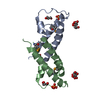
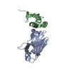
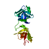
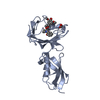
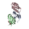
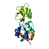
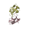
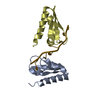
| Title | crystal structure of the eIF2 alpha subunit from saccharomyces cerevisia | ||||||
 Components Components | translation initiation factor 2 alpha subunit | ||||||
 Keywords Keywords | TRANSLATION / initiation factor / eIF2 / phosphorylation site | ||||||
| Function / homology |  Function and homology information Function and homology informationRecycling of eIF2:GDP / Cellular response to mitochondrial stress / ABC-family protein mediated transport / eukaryotic translation initiation factor 2 complex / multi-eIF complex / formation of translation preinitiation complex / eukaryotic 48S preinitiation complex / Formation of the ternary complex, and subsequently, the 43S complex / Translation initiation complex formation / Ribosomal scanning and start codon recognition ...Recycling of eIF2:GDP / Cellular response to mitochondrial stress / ABC-family protein mediated transport / eukaryotic translation initiation factor 2 complex / multi-eIF complex / formation of translation preinitiation complex / eukaryotic 48S preinitiation complex / Formation of the ternary complex, and subsequently, the 43S complex / Translation initiation complex formation / Ribosomal scanning and start codon recognition / L13a-mediated translational silencing of Ceruloplasmin expression / translation initiation factor activity / translational initiation / cytoplasmic stress granule / ribosome binding / ribosome / RNA binding / cytoplasm / cytosol Similarity search - Function | ||||||
| Biological species |  | ||||||
| Method |  X-RAY DIFFRACTION / MOLECULAR REPLACEMENT / Resolution: 2.86 Å X-RAY DIFFRACTION / MOLECULAR REPLACEMENT / Resolution: 2.86 Å | ||||||
 Authors Authors | Dhaliwal, S. / Hoffman, D.W. | ||||||
 Citation Citation | Journal: J.Mol.Biol. / Year: 2003 Title: The crystal structure of the N-terminal region of the alpha subunit of translation initiation factor 2 (eIF2alpha) from Saccharomyces cerevisiae provides a view of the loop containing serine ...Title: The crystal structure of the N-terminal region of the alpha subunit of translation initiation factor 2 (eIF2alpha) from Saccharomyces cerevisiae provides a view of the loop containing serine 51, the target of the eIF2alpha-specific kinases. Authors: Dhaliwal, S. / Hoffman, D.W. | ||||||
| History |
|
- Structure visualization
| Structure viewer | Molecule: MolmilJmol/JSmol |
|---|
- Downloads & links
-Download
| PDBx/mmCIF format | 1q46.cif.gz | 45.6 KB | Display | PDBx/mmCIF format |
|---|---|---|---|---|
| PDB format | pdb1q46.ent.gz | 33.3 KB | Display | PDB format |
| PDBx/mmJSON format | 1q46.json.gz | Tree view | PDBx/mmJSON format | |
| Others |  Other downloads Other downloads |
-Validation report
| Arichive directory | https://data.pdbj.org/pub/pdb/validation_reports/q4/1q46ftp://data.pdbj.org/pub/pdb/validation_reports/q4/1q46 | HTTPS FTP |
|---|
-Related structure data
| Similar structure data |
|---|

-Links
 PDBj
PDBj


- Assembly
| Deposited unit | 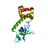
| ||||||||||
|---|---|---|---|---|---|---|---|---|---|---|---|
| 1 |
| ||||||||||
| Unit cell |
|
-Components
| #1: Protein | Mass: 20597.834 Da / Num. of mol.: 1 / Fragment: N-terminal two thirds of eIF2a / Source method: isolated from a natural source / Source: (natural) |
|---|
-Experimental details
-Experiment
| Experiment | Method: X-RAY DIFFRACTION / Number of used crystals: 1 |
|---|
- Sample preparation
| Crystal | Density Matthews: 4.35 Å3/Da / Density % sol: 71.75 % | |||||||||||||||
|---|---|---|---|---|---|---|---|---|---|---|---|---|---|---|---|---|
| Crystal grow | Temperature: 293 K / Method: vapor diffusion, hanging drop / pH: 4.6 Details: 0.1 M sodium acetate (pH=4.6), 1.8-2.5 M sodium formate, VAPOR DIFFUSION, HANGING DROP, temperature 293K | |||||||||||||||
| Crystal grow | *PLUS Method: unknown | |||||||||||||||
| Components of the solutions | *PLUS
|
-Data collection
| Diffraction | Mean temperature: 293 K |
|---|---|
| Diffraction source | Source: ROTATING ANODE / Type: RIGAKU RU200 / Wavelength: 1.54 Å |
| Detector | Type: MARRESEARCH / Detector: IMAGE PLATE / Date: Mar 20, 2003 / Details: osmium mirror |
| Radiation | Protocol: SINGLE WAVELENGTH / Monochromatic (M) / Laue (L): M / Scattering type: x-ray |
| Radiation wavelength | Wavelength: 1.54 Å / Relative weight: 1 |
| Reflection | Resolution: 2.86→20 Å / Num. all: 8830 / Num. obs: 8830 / % possible obs: 99.8 % / Observed criterion σ(F): 2 |
| Reflection shell | Resolution: 2.86→2.96 Å / Mean I/σ(I) obs: 1.3 / Rsym value: 0.583 / % possible all: 99.9 |
| Reflection | *PLUS Lowest resolution: 20 Å / Num. obs: 9935 / % possible obs: 99.5 % / Redundancy: 7.78 % / Num. measured all: 77383 / Rmerge(I) obs: 0.077 |
| Reflection shell | *PLUS Rmerge(I) obs: 0.583 |
- Processing
| Software |
| ||||||||||||||||||||
|---|---|---|---|---|---|---|---|---|---|---|---|---|---|---|---|---|---|---|---|---|---|
| Refinement | Method to determine structure: MOLECULAR REPLACEMENT Starting model: human eIF2 alpha subunit Resolution: 2.86→20 Å / σ(F): 0 / Stereochemistry target values: Engh & Huber
| ||||||||||||||||||||
| Refinement step | Cycle: LAST / Resolution: 2.86→20 Å
| ||||||||||||||||||||
| Refine LS restraints |
| ||||||||||||||||||||
| LS refinement shell | Resolution: 2.86→2.96 Å | ||||||||||||||||||||
| Refinement | *PLUS Lowest resolution: 20 Å / Rfactor Rfree: 0.243 / Rfactor Rwork: 0.222 | ||||||||||||||||||||
| Solvent computation | *PLUS | ||||||||||||||||||||
| Displacement parameters | *PLUS | ||||||||||||||||||||
| Refine LS restraints | *PLUS
|
