 Movie
Movie Controller

[English] 日本語
 Yorodumi
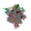
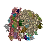
- PDB-4v6u: Promiscuous behavior of proteins in archaeal ribosomes revealed b... -
+ Open data

- Basic information
| Entry | Database: PDB / ID: 4v6u | |||||||||
|---|---|---|---|---|---|---|---|---|---|---|
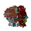
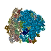
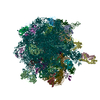
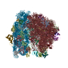
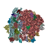
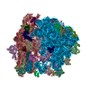
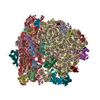
| Title | Promiscuous behavior of proteins in archaeal ribosomes revealed by cryo-EM: implications for evolution of eukaryotic ribosomes | |||||||||
 Components Components |
| |||||||||
 Keywords Keywords | RIBOSOME / archaea / archaeal / ribosomal / 70S / kink-turn / protein synthesis / RNA | |||||||||
| Function / homology |  Function and homology information Function and homology informationribonuclease P activity / tRNA 5'-leader removal / ribosomal large subunit biogenesis / maturation of LSU-rRNA from tricistronic rRNA transcript (SSU-rRNA, 5.8S rRNA, LSU-rRNA) / maturation of SSU-rRNA from tricistronic rRNA transcript (SSU-rRNA, 5.8S rRNA, LSU-rRNA) / maturation of SSU-rRNA / rRNA processing / regulation of translation / large ribosomal subunit / ribosomal small subunit assembly ...ribonuclease P activity / tRNA 5'-leader removal / ribosomal large subunit biogenesis / maturation of LSU-rRNA from tricistronic rRNA transcript (SSU-rRNA, 5.8S rRNA, LSU-rRNA) / maturation of SSU-rRNA from tricistronic rRNA transcript (SSU-rRNA, 5.8S rRNA, LSU-rRNA) / maturation of SSU-rRNA / rRNA processing / regulation of translation / large ribosomal subunit / ribosomal small subunit assembly / ribosomal small subunit biogenesis / 5S rRNA binding / ribosomal large subunit assembly / small ribosomal subunit / small ribosomal subunit rRNA binding / large ribosomal subunit rRNA binding / cytosolic small ribosomal subunit / cytosolic large ribosomal subunit / cytoplasmic translation / tRNA binding / negative regulation of translation / rRNA binding / structural constituent of ribosome / ribosome / translation / ribonucleoprotein complex / mRNA binding / RNA binding / zinc ion binding / cytosol / cytoplasm Similarity search - Function | |||||||||
| Biological species |   Pyrococcus furiosus (archaea) Pyrococcus furiosus (archaea) | |||||||||
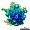
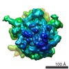
| Method | ELECTRON MICROSCOPY / single particle reconstruction / cryo EM / Resolution: 6.6 Å | |||||||||
 Authors Authors | Armache, J.-P. / Anger, A.M. / Marquez, V. / Frankenberg, S. / Froehlich, T. / Villa, E. / Berninghausen, O. / Thomm, M. / Arnold, G.J. / Beckmann, R. / Wilson, D.N. | |||||||||
 Citation Citation | Journal: Nucleic Acids Res / Year: 2013 Title: Promiscuous behaviour of archaeal ribosomal proteins: implications for eukaryotic ribosome evolution. Authors: Jean-Paul Armache / Andreas M Anger / Viter Márquez / Sibylle Franckenberg / Thomas Fröhlich / Elizabeth Villa / Otto Berninghausen / Michael Thomm / Georg J Arnold / Roland Beckmann / Daniel N Wilson /  Abstract: In all living cells, protein synthesis occurs on ribonucleoprotein particles called ribosomes. Molecular models have been reported for complete bacterial 70S and eukaryotic 80S ribosomes; however, ...In all living cells, protein synthesis occurs on ribonucleoprotein particles called ribosomes. Molecular models have been reported for complete bacterial 70S and eukaryotic 80S ribosomes; however, only molecular models of large 50S subunits have been reported for archaea. Here, we present a complete molecular model for the Pyrococcus furiosus 70S ribosome based on a 6.6 Å cryo-electron microscopy map. Moreover, we have determined cryo-electron microscopy reconstructions of the Euryarchaeota Methanococcus igneus and Thermococcus kodakaraensis 70S ribosomes and Crenarchaeota Staphylothermus marinus 50S subunit. Examination of these structures reveals a surprising promiscuous behavior of archaeal ribosomal proteins: We observe intersubunit promiscuity of S24e and L8e (L7ae), the latter binding to the head of the small subunit, analogous to S12e in eukaryotes. Moreover, L8e and L14e exhibit intrasubunit promiscuity, being present in two copies per archaeal 50S subunit, with the additional binding site of L14e analogous to the related eukaryotic r-protein L27e. Collectively, these findings suggest insights into the evolution of eukaryotic ribosomal proteins through increased copy number and binding site promiscuity. | |||||||||
| History |
|
- Structure visualization
| Movie |
Movie viewer |
|---|---|
| Structure viewer | Molecule: MolmilJmol/JSmol |
- Downloads & links
-Download
| PDBx/mmCIF format | 4v6u.cif.gz | 4 MB | Display | PDBx/mmCIF format |
|---|---|---|---|---|
| PDB format | pdb4v6u.ent.gz | Display | PDB format | |
| PDBx/mmJSON format | 4v6u.json.gz | Tree view | PDBx/mmJSON format | |
| Others |  Other downloads Other downloads |
-Validation report
| Arichive directory | https://data.pdbj.org/pub/pdb/validation_reports/v6/4v6uftp://data.pdbj.org/pub/pdb/validation_reports/v6/4v6u | HTTPS FTP |
|---|
-Related structure data
| Related structure data |  2009M 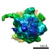 2170C 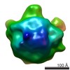 2171C 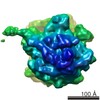 2172C M: map data used to model this data C: citing same article ( |
|---|---|
| Similar structure data |

-Links
 PDBj
PDBj


- Assembly
| Deposited unit | 
|
|---|---|
| 1 |
|
-Components
+30S ribosomal protein ... , 24 types, 25 molecules AQAKAIAGAWACABARADANAXAMAEAJAOAFASAYATAAAHAPAVB6AL
-Protein , 3 types, 3 molecules A9AUBk
| #9: Protein | Mass: 4868.993 Da / Num. of mol.: 1 / Fragment: SEE REMARK 999 / Source method: isolated from a natural source / Source: (natural) Pyrococcus furiosus (archaea) / Strain: ATCC 43587 / DSM 3638 / JCM 8422 / Vc1 |
|---|---|
| #30: Protein | Mass: 17394.236 Da / Num. of mol.: 1 / Source method: isolated from a natural source / Source: (natural) Pyrococcus furiosus (archaea) / Strain: ATCC 43587 / DSM 3638 / JCM 8422 / Vc1 / References: UniProt: Q8U0T4 |
| #43: Protein | Mass: 37166.969 Da / Num. of mol.: 1 / Source method: isolated from a natural source / Source: (natural) Pyrococcus furiosus (archaea) / Strain: ATCC 43587 / DSM 3638 / JCM 8422 / Vc1 / References: UniProt: Q8TZJ8 |
-RNA chain , 5 types, 5 molecules A1A2A0B1B3
| #11: RNA chain | Mass: 24880.865 Da / Num. of mol.: 1 / Source method: isolated from a natural source / Source: (natural) Pyrococcus furiosus (archaea) / Strain: ATCC 43587 / DSM 3638 / JCM 8422 / Vc1 |
|---|---|
| #21: RNA chain | Mass: 485455.500 Da / Num. of mol.: 1 / Source method: isolated from a natural source / Source: (natural) Pyrococcus furiosus (archaea) / Strain: ATCC 43587 / DSM 3638 / JCM 8422 / Vc1 |
| #27: RNA chain | Mass: 24538.561 Da / Num. of mol.: 1 / Source method: isolated from a natural source / Source: (natural) Pyrococcus furiosus (archaea) / Strain: ATCC 43587 / DSM 3638 / JCM 8422 / Vc1 |
| #67: RNA chain | Mass: 990820.375 Da / Num. of mol.: 1 / Source method: isolated from a natural source / Source: (natural) Pyrococcus furiosus (archaea) / Strain: ATCC 43587 / DSM 3638 / JCM 8422 / Vc1 |
| #68: RNA chain | Mass: 40689.262 Da / Num. of mol.: 1 / Source method: isolated from a natural source / Source: (natural) Pyrococcus furiosus (archaea) / Strain: ATCC 43587 / DSM 3638 / JCM 8422 / Vc1 |
+50S ribosomal protein ... , 36 types, 39 molecules A3BGB4BYBOBCB5BKBLBfBUBbBeBEBaBTBWBiBABIBRBQBVBjBBBDBFBhBHBZ...
-Details
| Sequence details | THE SEQUENCE OF 30S RIBOSOMAL PROTEIN SX IS NOT KNOWN. |
|---|
-Experimental details
-Experiment
| Experiment | Method: ELECTRON MICROSCOPY |
|---|---|
| EM experiment | Aggregation state: PARTICLE / 3D reconstruction method: single particle reconstruction |
- Sample preparation
| Component | Name: Pyrococcus furiosus 70S ribosome / Type: RIBOSOME |
|---|---|
| Buffer solution | Name: 56 mM Tris pH 8.2, 250 mM KOAc, 80 mM NH4OAc, 50 mM MgCl2, 1 mM DTT, 2 mM ADPNP pH: 8.2 Details: 56 mM Tris pH 8.2, 250 mM KOAc, 80 mM NH4OAc, 50 mM MgCl2, 1 mM DTT, 2 mM ADPNP |
| Specimen | Embedding applied: NO / Shadowing applied: NO / Staining applied: NO / Vitrification applied: YES |
| Specimen support | Details: Quantifoil grids (3/3) with 2 nm carbon on top |
| Vitrification | Instrument: FEI VITROBOT MARK I / Cryogen name: ETHANE / Humidity: 95 % Details: Blot for 10 seconds before plunging into liquid ethane, using 2 layers of filter paper (FEI VITROBOT) |
- Electron microscopy imaging
| Experimental equipment |  Model: Titan Krios / Image courtesy: FEI Company |
|---|---|
| Microscopy | Model: FEI TITAN KRIOS |
| Electron gun | Electron source:  FIELD EMISSION GUN / Accelerating voltage: 200 kV / Illumination mode: FLOOD BEAM FIELD EMISSION GUN / Accelerating voltage: 200 kV / Illumination mode: FLOOD BEAM |
| Electron lens | Mode: BRIGHT FIELD / Nominal magnification: 75000 X / Nominal defocus max: 3600 nm / Nominal defocus min: 1200 nm / Cs: 2.7 mm |
| Image recording | Electron dose: 25 e/Å2 / Film or detector model: TVIPS TEMCAM-F416 (4k x 4k) |
| Radiation | Protocol: SINGLE WAVELENGTH / Monochromatic (M) / Laue (L): M / Scattering type: x-ray |
| Radiation wavelength | Relative weight: 1 |
- Processing
| EM software | Name: SPIDER / Category: 3D reconstruction | ||||||||||||
|---|---|---|---|---|---|---|---|---|---|---|---|---|---|
| CTF correction | Details: Wiener Filter | ||||||||||||
| Symmetry | Point symmetry: C1 (asymmetric) | ||||||||||||
| 3D reconstruction | Method: Single Particle / Resolution: 6.6 Å / Resolution method: FSC 0.5 CUT-OFF / Num. of particles: 10000 / Nominal pixel size: 1.24 Å / Actual pixel size: 1.24 Å Details: sorting for ribosome conformation and ligand presence was performed Symmetry type: POINT | ||||||||||||
| Refinement step | Cycle: LAST
|
