 Movie
Movie Controller

[English] 日本語
 Yorodumi
- PDB-4v4a: Crystal Structure of the Wild Type Ribosome from E. Coli 70S Ribosome. -
+ Open data

- Basic information
| Entry | Database: PDB / ID: 4v4a | |||||||||
|---|---|---|---|---|---|---|---|---|---|---|
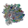
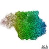
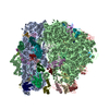
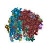
| Title | Crystal Structure of the Wild Type Ribosome from E. Coli 70S Ribosome. | |||||||||
 Components Components |
| |||||||||
 Keywords Keywords | RIBOSOME / 30S RIBOSOMAL SUBUNIT / PROTEIN-PROTEIN COMPLEX / PROTEIN-RNA COMPLEX / RNA-RNA COMPLEX | |||||||||
| Function / homology | RNA / RNA (> 10) / RNA (> 100) / RNA (> 1000) Function and homology information Function and homology information | |||||||||
| Biological species |  | |||||||||
| Method |  X-RAY DIFFRACTION / SYNCHROTRON / MOLECULAR REPLACEMENT / Resolution: 9.5 Å X-RAY DIFFRACTION / SYNCHROTRON / MOLECULAR REPLACEMENT / Resolution: 9.5 Å | |||||||||
 Authors Authors | Vila-Sanjurjo, A. / Ridgeway, W.K. / Seymaner, V. / Zhang, W. / Santoso, S. / Yu, K. / Cate, J.H.D. | |||||||||
 Citation Citation | Journal: Proc.Natl.Acad.Sci.USA / Year: 2003 Title: X-ray crystal structures of the WT and a hyper-accurate ribosome from Escherichia coli Authors: Vila-Sanjurjo, A. / Ridgeway, W.K. / Seymaner, V. / Zhang, W. / Santoso, S. / Yu, K. / Cate, J.H.D. | |||||||||
| History |
| |||||||||
| Remark 400 | COMPOUND PDB ENTRIES 1PNX AND 1PNY REPRESENT ONE CRYSTAL STRUCTURE OF THE E. COLI 70S RIBOSOME. ...COMPOUND PDB ENTRIES 1PNX AND 1PNY REPRESENT ONE CRYSTAL STRUCTURE OF THE E. COLI 70S RIBOSOME. THIS FILE, 1PNX, CONTAINS ONLY MOLECULES OF THE 30S RIBOSOMAL SUBUNIT. THE 50S SUBUNIT IS IN THE PDB FILE 1PNY. | |||||||||
| Remark 999 | SEQUENCE The 30S subunit is derived mainly from 1J5E, the T. thermophilus 30S subunit, with ...SEQUENCE The 30S subunit is derived mainly from 1J5E, the T. thermophilus 30S subunit, with modifications to the rRNA to make it match E. coli insertions and deletions. The sequence of this subunit represent that of pdb entry, 1J5E. As a result, no dbref was provided. |
- Structure visualization
| Structure viewer | Molecule: MolmilJmol/JSmol |
|---|
- Downloads & links
-Download
| PDBx/mmCIF format | 4v4a.cif.gz | 2.6 MB | Display | PDBx/mmCIF format |
|---|---|---|---|---|
| PDB format | pdb4v4a.ent.gz | Display | PDB format | |
| PDBx/mmJSON format | 4v4a.json.gz | Tree view | PDBx/mmJSON format | |
| Others |  Other downloads Other downloads |
-Validation report
| Arichive directory | https://data.pdbj.org/pub/pdb/validation_reports/v4/4v4aftp://data.pdbj.org/pub/pdb/validation_reports/v4/4v4a | HTTPS FTP |
|---|
-Related structure data
| Related structure data |  4v49C  1j5eS  1lnr C: citing same article ( S: Starting model for refinement |
|---|---|
| Similar structure data |

-Links
 PDBj
PDBj


- Assembly
| Deposited unit | 
| ||||||||
|---|---|---|---|---|---|---|---|---|---|
| 1 |
| ||||||||
| Unit cell |
|
-Components
-RNA chain , 3 types, 3 molecules AAB0B9
| #1: RNA chain | Mass: 498797.375 Da / Num. of mol.: 1 / Source method: isolated from a natural source / Source: (natural) |
|---|---|
| #21: RNA chain | Mass: 935779.625 Da / Num. of mol.: 1 / Source method: isolated from a natural source / Source: (natural) |
| #22: RNA chain | Mass: 38029.777 Da / Num. of mol.: 1 / Source method: isolated from a natural source / Source: (natural) |
-30S ribosomal protein ... , 19 types, 19 molecules ABACADAEAFAGAHAIAJAKALAMANAOAPAQARASAT
| #2: Protein | Mass: 26987.271 Da / Num. of mol.: 1 / Source method: isolated from a natural source / Source: (natural) |
|---|---|
| #3: Protein | Mass: 22862.430 Da / Num. of mol.: 1 / Source method: isolated from a natural source / Source: (natural) |
| #4: Protein | Mass: 24242.254 Da / Num. of mol.: 1 / Source method: isolated from a natural source / Source: (natural) |
| #5: Protein | Mass: 16331.079 Da / Num. of mol.: 1 / Source method: isolated from a natural source / Source: (natural) |
| #6: Protein | Mass: 11988.753 Da / Num. of mol.: 1 / Source method: isolated from a natural source / Source: (natural) |
| #7: Protein | Mass: 17919.775 Da / Num. of mol.: 1 / Source method: isolated from a natural source / Source: (natural) |
| #8: Protein | Mass: 15868.570 Da / Num. of mol.: 1 / Source method: isolated from a natural source / Source: (natural) |
| #9: Protein | Mass: 14298.466 Da / Num. of mol.: 1 / Source method: isolated from a natural source / Source: (natural) |
| #10: Protein | Mass: 11299.176 Da / Num. of mol.: 1 / Source method: isolated from a natural source / Source: (natural) |
| #11: Protein | Mass: 12606.369 Da / Num. of mol.: 1 / Source method: isolated from a natural source / Source: (natural) |
| #12: Protein | Mass: 13804.311 Da / Num. of mol.: 1 / Source method: isolated from a natural source / Source: (natural) |
| #13: Protein | Mass: 14207.666 Da / Num. of mol.: 1 / Source method: isolated from a natural source / Source: (natural) |
| #14: Protein | Mass: 7027.529 Da / Num. of mol.: 1 / Source method: isolated from a natural source / Source: (natural) |
| #15: Protein | Mass: 10447.213 Da / Num. of mol.: 1 / Source method: isolated from a natural source / Source: (natural) |
| #16: Protein | Mass: 9924.469 Da / Num. of mol.: 1 / Source method: isolated from a natural source / Source: (natural) |
| #17: Protein | Mass: 12193.475 Da / Num. of mol.: 1 / Source method: isolated from a natural source / Source: (natural) |
| #18: Protein | Mass: 8483.172 Da / Num. of mol.: 1 / Source method: isolated from a natural source / Source: (natural) |
| #19: Protein | Mass: 9203.743 Da / Num. of mol.: 1 / Source method: isolated from a natural source / Source: (natural) |
| #20: Protein | Mass: 10907.060 Da / Num. of mol.: 1 / Source method: isolated from a natural source / Source: (natural) |
+50S ribosomal protein ... , 30 types, 30 molecules BABBBCBDBEBFBGBHBIBJBKBLBMBNBOBPBQBRBSBUBVBWBXBYBZB1B2B3B4B5
-Protein , 1 types, 1 molecules BT
| #42: Protein | Mass: 18929.711 Da / Num. of mol.: 1 / Source method: isolated from a natural source / Source: (natural) |
|---|
-Details
| Has protein modification | N |
|---|
-Experimental details
-Experiment
| Experiment | Method: X-RAY DIFFRACTION / Number of used crystals: 10 |
|---|
- Sample preparation
| Crystal | Density Matthews: 5.28 Å3/Da / Density % sol: 76.7 % | ||||||||||||||||||||||||||||||||||||||||||||||||||||
|---|---|---|---|---|---|---|---|---|---|---|---|---|---|---|---|---|---|---|---|---|---|---|---|---|---|---|---|---|---|---|---|---|---|---|---|---|---|---|---|---|---|---|---|---|---|---|---|---|---|---|---|---|---|
| Crystal grow | Temperature: 277 K / Method: vapor diffusion / pH: 6.5 Details: MPD, ethanol, magnesium chloride, ammonium chloride, spermine, spermidine, MES , pH 6.5, VAPOR DIFFUSION, temperature 277K | ||||||||||||||||||||||||||||||||||||||||||||||||||||
| Components of the solutions |
|
-Data collection
| Diffraction | Mean temperature: 110 K | |||||||||
|---|---|---|---|---|---|---|---|---|---|---|
| Diffraction source | Source: SYNCHROTRON / Site: ALS  / Beamline: 5.0.2 / Wavelength: 1.0, 1.1 / Beamline: 5.0.2 / Wavelength: 1.0, 1.1 | |||||||||
| Detector | Type: ADSC QUANTUM 4 / Detector: CCD / Date: Dec 8, 2000 | |||||||||
| Radiation | Monochromator: Si 111 / Protocol: SINGLE WAVELENGTH / Monochromatic (M) / Laue (L): M / Scattering type: x-ray | |||||||||
| Radiation wavelength |
| |||||||||
| Reflection | Resolution: 9.5→300 Å / Num. all: 29028 / Num. obs: 26821 / % possible obs: 92.1 % / Observed criterion σ(F): 0 / Observed criterion σ(I): -3 / Redundancy: 25.1 % / Rmerge(I) obs: 0.142 / Net I/σ(I): 44.8 | |||||||||
| Reflection shell | Resolution: 9.5→9.68 Å / Redundancy: 9.7 % / Rmerge(I) obs: 0.549 / Mean I/σ(I) obs: 2.5 / % possible all: 71.8 |
- Processing
| Software |
| |||||||||||||||||||||||||
|---|---|---|---|---|---|---|---|---|---|---|---|---|---|---|---|---|---|---|---|---|---|---|---|---|---|---|
| Refinement | Method to determine structure: MOLECULAR REPLACEMENT Starting model: 1J5E,1LNR Resolution: 9.5→70 Å / Isotropic thermal model: ANISOTROPIC / Cross valid method: THROUGHOUT / σ(F): 0 / Stereochemistry target values: NA Details: 87 RIGID-BODIES REFINEMENT, GROUPED B-FACTOR REFINEMENT. There are many long O3'-P distances. Coordinates used for solving and refining this structure mainly comes from PDB entry, 1J5E, ...Details: 87 RIGID-BODIES REFINEMENT, GROUPED B-FACTOR REFINEMENT. There are many long O3'-P distances. Coordinates used for solving and refining this structure mainly comes from PDB entry, 1J5E, where the sequence represents that of T. thermophilus.
| |||||||||||||||||||||||||
| Displacement parameters | Biso mean: 696.1 Å2 | |||||||||||||||||||||||||
| Refinement step | Cycle: LAST / Resolution: 9.5→70 Å
|
