 Movie
Movie Controller

[English] 日本語
 Yorodumi
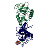
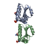
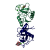
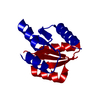
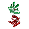
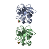
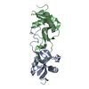
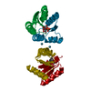
- PDB-4uso: X-ray structure of the CCL2 lectin in complex with sialyl lewis X -
+ Open data

- Basic information
| Entry | Database: PDB / ID: 4uso | |||||||||
|---|---|---|---|---|---|---|---|---|---|---|
| Title | X-ray structure of the CCL2 lectin in complex with sialyl lewis X | |||||||||
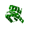
 Components Components | CCL2 LECTIN | |||||||||
 Keywords Keywords | SUGAR BINDING PROTEIN / DIMERIC / FUNGAL | |||||||||
| Function / homology |  Function and homology information Function and homology information | |||||||||
| Biological species |  COPRINOPSIS CINEREA (fungus) COPRINOPSIS CINEREA (fungus) | |||||||||
| Method |  X-RAY DIFFRACTION / SYNCHROTRON / MOLECULAR REPLACEMENT / Resolution: 1.95 Å X-RAY DIFFRACTION / SYNCHROTRON / MOLECULAR REPLACEMENT / Resolution: 1.95 Å | |||||||||
 Authors Authors | Bleuler-Martinez, S. / Varrot, A. / Schubert, M. / Stutz, M. / Sieber, R. / Hengartner, M. / Aebi, M. / Kunzler, M. | |||||||||
 Citation Citation | Journal: Glycobiology / Year: 2017 Title: Dimerization of the fungal defense lectin CCL2 is essential for its toxicity against nematodes. Authors: Bleuler-Martinez, S. / Stutz, K. / Sieber, R. / Collot, M. / Mallet, J.M. / Hengartner, M. / Schubert, M. / Varrot, A. / Kunzler, M. | |||||||||
| History |
| |||||||||
| Remark 700 | SHEET DETERMINATION METHOD: DSSP THE SHEETS PRESENTED AS "AA" IN EACH CHAIN ON SHEET RECORDS BELOW ... SHEET DETERMINATION METHOD: DSSP THE SHEETS PRESENTED AS "AA" IN EACH CHAIN ON SHEET RECORDS BELOW IS ACTUALLY AN 6-STRANDED BARREL THIS IS REPRESENTED BY A 7-STRANDED SHEET IN WHICH THE FIRST AND LAST STRANDS ARE IDENTICAL. THE SHEETS PRESENTED AS "BA" IN EACH CHAIN ON SHEET RECORDS BELOW IS ACTUALLY AN 6-STRANDED BARREL THIS IS REPRESENTED BY A 7-STRANDED SHEET IN WHICH THE FIRST AND LAST STRANDS ARE IDENTICAL. THE SHEETS PRESENTED AS "CA" IN EACH CHAIN ON SHEET RECORDS BELOW IS ACTUALLY AN 6-STRANDED BARREL THIS IS REPRESENTED BY A 7-STRANDED SHEET IN WHICH THE FIRST AND LAST STRANDS ARE IDENTICAL. |
- Structure visualization
| Structure viewer | Molecule: MolmilJmol/JSmol |
|---|
- Downloads & links
-Download
| PDBx/mmCIF format | 4uso.cif.gz | 219.8 KB | Display | PDBx/mmCIF format |
|---|---|---|---|---|
| PDB format | pdb4uso.ent.gz | 177.7 KB | Display | PDB format |
| PDBx/mmJSON format | 4uso.json.gz | Tree view | PDBx/mmJSON format | |
| Others |  Other downloads Other downloads |
-Validation report
| Arichive directory | https://data.pdbj.org/pub/pdb/validation_reports/us/4usoftp://data.pdbj.org/pub/pdb/validation_reports/us/4uso | HTTPS FTP |
|---|
-Related structure data
| Related structure data |  4uspC  2lieS C: citing same article ( S: Starting model for refinement |
|---|---|
| Similar structure data |


-Links
 PDBj
PDBj

- Assembly
| Deposited unit | 
| ||||||||||||||||
|---|---|---|---|---|---|---|---|---|---|---|---|---|---|---|---|---|---|
| 1 | 
| ||||||||||||||||
| 2 | 
| ||||||||||||||||
| Unit cell |
| ||||||||||||||||
| Components on special symmetry positions |
| ||||||||||||||||
| Noncrystallographic symmetry (NCS) | NCS oper:
|
-Components
| #1: Protein | Mass: 16604.281 Da / Num. of mol.: 4 Source method: isolated from a genetically manipulated source Source: (gene. exp.) COPRINOPSIS CINEREA (fungus) / Production host:  #2: Polysaccharide | N-acetyl-alpha-neuraminic acid-(2-3)-beta-D-galactopyranose-(1-4)-[alpha-L-fucopyranose-(1-3)]2- ...N-acetyl-alpha-neuraminic acid-(2-3)-beta-D-galactopyranose-(1-4)-[alpha-L-fucopyranose-(1-3)]2-acetamido-2-deoxy-beta-D-glucopyranose / Sialyl-Lewis X antigen / beta anomer | 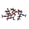  Source method: isolated from a genetically manipulated source Details: oligosaccharide with branches / References: Sialyl-Lewis X antigen, beta anomer #3: Water | ChemComp-HOH / |  Mass: 18.015 Da / Num. of mol.: 229 / Source method: isolated from a natural source / Formula: H2O Mass: 18.015 Da / Num. of mol.: 229 / Source method: isolated from a natural source / Formula: H2OSequence details | THE FIRST 12 AMINO ACIDS ARE FROM THE NTERMINAL HISTAG | |
|---|
-Experimental details
-Experiment
| Experiment | Method: X-RAY DIFFRACTION / Number of used crystals: 1 |
|---|
- Sample preparation
| Crystal | Density Matthews: 2.34 Å3/Da / Density % sol: 47.42 % / Description: NONE |
|---|---|
| Crystal grow | pH: 8.5 Details: MIDAS 2-28. 30% GLYCEROL ETHOXYLATE AND 0.1M TRIS PH 8.5 |
-Data collection
| Diffraction | Mean temperature: 100 K |
|---|---|
| Diffraction source | Source: SYNCHROTRON / Site: ESRF  / Beamline: ID23-1 / Wavelength: 0.9732 / Beamline: ID23-1 / Wavelength: 0.9732 |
| Detector | Type: DECTRIS PILATUS / Detector: PIXEL / Date: Sep 25, 2013 |
| Radiation | Protocol: SINGLE WAVELENGTH / Monochromatic (M) / Laue (L): M / Scattering type: x-ray |
| Radiation wavelength | Wavelength: 0.9732 Å / Relative weight: 1 |
| Reflection | Resolution: 1.95→29.23 Å / Num. obs: 46087 / % possible obs: 99.2 % / Observed criterion σ(I): 2 / Redundancy: 6.3 % / Rmerge(I) obs: 0.05 / Net I/σ(I): 18 |
| Reflection shell | Resolution: 1.95→2 Å / Redundancy: 6.6 % / Rmerge(I) obs: 0.42 / Mean I/σ(I) obs: 4.4 / % possible all: 100 |
- Processing
| Software |
| ||||||||||||||||||||||||||||||||||||||||||||||||||||||||||||||||||||||||||||||||||||||||||||||||||||||||||||||||||||||||||||||||||||||||||||||||||||||||||||||||||||||||||||||||||||||
|---|---|---|---|---|---|---|---|---|---|---|---|---|---|---|---|---|---|---|---|---|---|---|---|---|---|---|---|---|---|---|---|---|---|---|---|---|---|---|---|---|---|---|---|---|---|---|---|---|---|---|---|---|---|---|---|---|---|---|---|---|---|---|---|---|---|---|---|---|---|---|---|---|---|---|---|---|---|---|---|---|---|---|---|---|---|---|---|---|---|---|---|---|---|---|---|---|---|---|---|---|---|---|---|---|---|---|---|---|---|---|---|---|---|---|---|---|---|---|---|---|---|---|---|---|---|---|---|---|---|---|---|---|---|---|---|---|---|---|---|---|---|---|---|---|---|---|---|---|---|---|---|---|---|---|---|---|---|---|---|---|---|---|---|---|---|---|---|---|---|---|---|---|---|---|---|---|---|---|---|---|---|---|---|
| Refinement | Method to determine structure: MOLECULAR REPLACEMENT Starting model: PDB ENTRY 2LIE Resolution: 1.95→29.85 Å / Cor.coef. Fo:Fc: 0.964 / Cor.coef. Fo:Fc free: 0.948 / SU B: 5.773 / SU ML: 0.087 / Cross valid method: THROUGHOUT / ESU R: 0.14 / ESU R Free: 0.127 / Stereochemistry target values: MAXIMUM LIKELIHOOD Details: HYDROGENS HAVE BEEN ADDED IN THE RIDING POSITIONS. U VALUES WITH TLS ADDED LOCAL NCS DISORDERED REGIONS WERE MODELED STEREOCHEMICALLY
| ||||||||||||||||||||||||||||||||||||||||||||||||||||||||||||||||||||||||||||||||||||||||||||||||||||||||||||||||||||||||||||||||||||||||||||||||||||||||||||||||||||||||||||||||||||||
| Solvent computation | Ion probe radii: 0.8 Å / Shrinkage radii: 0.8 Å / VDW probe radii: 1.2 Å / Solvent model: MASK | ||||||||||||||||||||||||||||||||||||||||||||||||||||||||||||||||||||||||||||||||||||||||||||||||||||||||||||||||||||||||||||||||||||||||||||||||||||||||||||||||||||||||||||||||||||||
| Displacement parameters | Biso mean: 48.158 Å2
| ||||||||||||||||||||||||||||||||||||||||||||||||||||||||||||||||||||||||||||||||||||||||||||||||||||||||||||||||||||||||||||||||||||||||||||||||||||||||||||||||||||||||||||||||||||||
| Refinement step | Cycle: LAST / Resolution: 1.95→29.85 Å
| ||||||||||||||||||||||||||||||||||||||||||||||||||||||||||||||||||||||||||||||||||||||||||||||||||||||||||||||||||||||||||||||||||||||||||||||||||||||||||||||||||||||||||||||||||||||
| Refine LS restraints |
|