 Movie
Movie Controller

[English] 日本語
 Yorodumi
- PDB-1rdl: MANNOSE-BINDING PROTEIN, SUBTILISIN DIGEST FRAGMENT COMPLEX WITH ... -
+ Open data

- Basic information
| Entry | Database: PDB / ID: 1rdl | ||||||
|---|---|---|---|---|---|---|---|
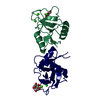
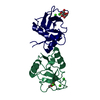
| Title | MANNOSE-BINDING PROTEIN, SUBTILISIN DIGEST FRAGMENT COMPLEX WITH ALPHA-METHYL-D-MANNOPYRANOSIDE (0.2 M) | ||||||
 Components Components | MANNOSE-BINDING PROTEIN-C | ||||||
 Keywords Keywords | LECTIN / C-TYPE LECTIN / CALCIUM-BINDING PROTEIN | ||||||
| Function / homology |  Function and homology information Function and homology informationLectin pathway of complement activation / positive regulation of opsonization / negative regulation of viral process / positive regulation of protein processing / positive regulation of complement activation / complement activation, lectin pathway / galactose binding / surfactant homeostasis / cell surface pattern recognition receptor signaling pathway / symbiont cell surface ...Lectin pathway of complement activation / positive regulation of opsonization / negative regulation of viral process / positive regulation of protein processing / positive regulation of complement activation / complement activation, lectin pathway / galactose binding / surfactant homeostasis / cell surface pattern recognition receptor signaling pathway / symbiont cell surface / phosphatidylinositol-4-phosphate binding / antibacterial innate immune response / D-mannose binding / pattern recognition receptor activity / multivesicular body / positive regulation of phagocytosis / antiviral innate immune response / calcium-dependent protein binding / protease binding / innate immune response / defense response to Gram-positive bacterium / signaling receptor binding / calcium ion binding / cell surface / protein-containing complex / proteolysis / extracellular region / identical protein binding Similarity search - Function | ||||||
| Biological species |   Rattus rattus (black rat) Rattus rattus (black rat) | ||||||
| Method |  X-RAY DIFFRACTION / Resolution: 1.7 Å X-RAY DIFFRACTION / Resolution: 1.7 Å | ||||||
 Authors Authors | Ng, K.K.-S. / Drickamer, K. / Weis, W.I. | ||||||
 Citation Citation | Journal: J.Biol.Chem. / Year: 1996 Title: Structural analysis of monosaccharide recognition by rat liver mannose-binding protein. Authors: Ng, K.K. / Drickamer, K. / Weis, W.I. #1: Journal: Nature / Year: 1992Title: Structure of a C-Type Mannose-Binding Protein Complexed with an Oligosaccharide Authors: Weis, W.I. / Drickamer, K. / Hendrickson, W.A. #2: Journal: Science / Year: 1991Title: Structure of the Calcium-Dependent Lectin Domain from a Rat Mannose-Binding Protein Determined by MAD Phasing Authors: Weis, W.I. / Kahn, R. / Fourme, R. / Drickamer, K. / Hendrickson, W.A. #3: Journal: J.Biol.Chem. / Year: 1991Title: Physical Characterization and Crystallization of the Carbohydrate-Recognition Domain of a Mannose-Binding Protein from Rat Authors: Weis, W.I. / Crichlow, G.V. / Murthy, H.M.K. / Hendrickson, W.A. / Drickamer, K. #4: Journal: J.Biol.Chem. / Year: 1990Title: Differential Recognition of Core and Terminal Portions of Oligosaccharide Ligands by Carbohydrate-Recognition Domains of Two Mannose-Binding Proteins Authors: Childs, R.A. / Feizi, T. / Yuen, C.-T. / Drickamer, K. / Quesenberry, M.S. | ||||||
| History |
|
- Structure visualization
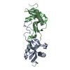
| Structure viewer | Molecule: MolmilJmol/JSmol |
|---|
- Downloads & links
-Download
| PDBx/mmCIF format | 1rdl.cif.gz | 67 KB | Display | PDBx/mmCIF format |
|---|---|---|---|---|
| PDB format | pdb1rdl.ent.gz | 48.1 KB | Display | PDB format |
| PDBx/mmJSON format | 1rdl.json.gz | Tree view | PDBx/mmJSON format | |
| Others |  Other downloads Other downloads |
-Validation report
| Arichive directory | https://data.pdbj.org/pub/pdb/validation_reports/rd/1rdlftp://data.pdbj.org/pub/pdb/validation_reports/rd/1rdl | HTTPS FTP |
|---|
-Related structure data
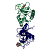
| Related structure data | 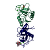 1rdiC 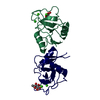 1rdjC  1rdkC 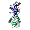 1rdmC  1rdnC  1rdoC C: citing same article ( |
|---|---|
| Similar structure data |




-Links
 PDBj
PDBj

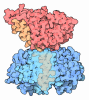
- Assembly
| Deposited unit | 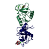
| ||||||||
|---|---|---|---|---|---|---|---|---|---|
| 1 |
| ||||||||
| Unit cell |
| ||||||||
| Atom site foot note | 1: CIS PROLINE - PRO 1 191 / 2: CIS PROLINE - PRO 2 191 | ||||||||
| Noncrystallographic symmetry (NCS) | NCS oper: (Code: given Matrix: (0.999843, 0.016055, 0.007514), Vector: Details | MTRIX THE TRANSFORMATIONS PRESENTED ON MTRIX RECORDS BELOW DESCRIBE NON-CRYSTALLOGRAPHIC RELATIONSHIPS AMONG THE VARIOUS DOMAINS IN THIS ENTRY. APPLYING THE APPROPRIATE MTRIX TRANSFORMATION TO THE RESIDUES LISTED FIRST WILL YIELD APPROXIMATE COORDINATES FOR THE RESIDUES LISTED SECOND. APPLIED TO TRANSFORMED TO MTRIX RESIDUES RESIDUES RMSD M1 1 115 .. 1 225 2 115 .. 2 225 0.307 | |
-Components
| #1: Protein | Mass: 12675.127 Da / Num. of mol.: 2 / Fragment: SUBTILISIN FRAGMENT (RESIDUES 114 - 226) Source method: isolated from a genetically manipulated source Source: (gene. exp.) Rattus rattus (black rat)Description: THE BACTERIALLY EXPRESSED MATERIAL IS DIGESTED WITH SUBTILISIN TO PRODUCE THE PROTEIN USED IN THE CRYSTAL STRUCTURE ANALYSIS Organ: LIVER / Plasmid: PINIIIOMPA2 / Production host:   Escherichia coli (E. coli) / References: UniProt: P08661 Escherichia coli (E. coli) / References: UniProt: P08661#2: Sugar |  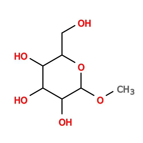 Type: D-saccharide / Mass: 194.182 Da / Num. of mol.: 2 Type: D-saccharide / Mass: 194.182 Da / Num. of mol.: 2Source method: isolated from a genetically manipulated source Formula: C7H14O6 #3: Chemical | ChemComp-CA /   Mass: 40.078 Da / Num. of mol.: 4 / Source method: obtained synthetically / Formula: Ca Mass: 40.078 Da / Num. of mol.: 4 / Source method: obtained synthetically / Formula: Ca#4: Chemical | ChemComp-CL / |   Mass: 35.453 Da / Num. of mol.: 1 / Source method: obtained synthetically / Formula: Cl Mass: 35.453 Da / Num. of mol.: 1 / Source method: obtained synthetically / Formula: Cl#5: Water | ChemComp-HOH / |  Mass: 18.015 Da / Num. of mol.: 297 / Source method: isolated from a natural source / Formula: H2O Mass: 18.015 Da / Num. of mol.: 297 / Source method: isolated from a natural source / Formula: H2OHas protein modification | Y | |
|---|
-Experimental details
-Experiment
| Experiment | Method: X-RAY DIFFRACTION / Number of used crystals: 1 |
|---|
- Sample preparation
| Crystal | Density Matthews: 2.59 Å3/Da / Density % sol: 52.51 % | ||||||||||||||||||||||||||||||||||||||||||||||||||||||||||||||||||||||||
|---|---|---|---|---|---|---|---|---|---|---|---|---|---|---|---|---|---|---|---|---|---|---|---|---|---|---|---|---|---|---|---|---|---|---|---|---|---|---|---|---|---|---|---|---|---|---|---|---|---|---|---|---|---|---|---|---|---|---|---|---|---|---|---|---|---|---|---|---|---|---|---|---|---|
| Crystal grow | pH: 7.4 / Details: pH 7.4 | ||||||||||||||||||||||||||||||||||||||||||||||||||||||||||||||||||||||||
| Crystal grow | *PLUS Temperature: 22 ℃ / Method: vapor diffusion, hanging drop | ||||||||||||||||||||||||||||||||||||||||||||||||||||||||||||||||||||||||
| Components of the solutions | *PLUS
|
-Data collection
| Diffraction | Mean temperature: 100 K |
|---|---|
| Diffraction source | Wavelength: 1.5418 Å |
| Detector | Type: RIGAKU / Detector: IMAGE PLATE / Date: Jun 4, 1994 |
| Radiation | Monochromator: GRAPHITE / Monochromatic (M) / Laue (L): M / Scattering type: x-ray |
| Radiation wavelength | Wavelength: 1.5418 Å / Relative weight: 1 |
| Reflection | Resolution: 1.7→10 Å / Num. obs: 28080 / % possible obs: 94.5 % / Observed criterion σ(I): 0 / Redundancy: 3.2 % / Rmerge(I) obs: 0.045 |
| Reflection | *PLUS Rmerge(I) obs: 0.045 |
- Processing
| Software |
| ||||||||||||||||||||||||||||||||||||||||||||||||||||||||||||||||||||||||||||||||
|---|---|---|---|---|---|---|---|---|---|---|---|---|---|---|---|---|---|---|---|---|---|---|---|---|---|---|---|---|---|---|---|---|---|---|---|---|---|---|---|---|---|---|---|---|---|---|---|---|---|---|---|---|---|---|---|---|---|---|---|---|---|---|---|---|---|---|---|---|---|---|---|---|---|---|---|---|---|---|---|---|---|
| Refinement | Resolution: 1.7→10 Å / σ(F): 2
| ||||||||||||||||||||||||||||||||||||||||||||||||||||||||||||||||||||||||||||||||
| Displacement parameters | Biso mean: 21.4 Å2 | ||||||||||||||||||||||||||||||||||||||||||||||||||||||||||||||||||||||||||||||||
| Refinement step | Cycle: LAST / Resolution: 1.7→10 Å
| ||||||||||||||||||||||||||||||||||||||||||||||||||||||||||||||||||||||||||||||||
| Refine LS restraints |
| ||||||||||||||||||||||||||||||||||||||||||||||||||||||||||||||||||||||||||||||||
| Software | *PLUS Name: X-PLOR / Classification: refinement | ||||||||||||||||||||||||||||||||||||||||||||||||||||||||||||||||||||||||||||||||
| Refinement | *PLUS | ||||||||||||||||||||||||||||||||||||||||||||||||||||||||||||||||||||||||||||||||
| Solvent computation | *PLUS | ||||||||||||||||||||||||||||||||||||||||||||||||||||||||||||||||||||||||||||||||
| Displacement parameters | *PLUS | ||||||||||||||||||||||||||||||||||||||||||||||||||||||||||||||||||||||||||||||||
| Refine LS restraints | *PLUS
|
