 Movie
Movie Controller


+ Open data

- Basic information
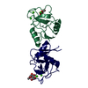
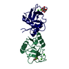
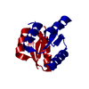
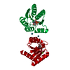
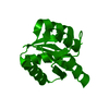
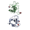
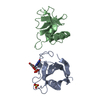
| Entry | Database: PDB / ID: 1kzb | ||||||
|---|---|---|---|---|---|---|---|
| Title | Complex of MBP-C and trimannosyl core | ||||||
 Components Components | MANNOSE-BINDING PROTEIN C | ||||||
 Keywords Keywords | IMMUNE SYSTEM / SUGAR BINDING PROTEIN / protein-carbohydrate complex | ||||||
| Function / homology |  Function and homology information Function and homology informationLectin pathway of complement activation / Initial triggering of complement / activation of membrane attack complex / positive regulation of opsonization / positive regulation of complement activation / complement activation, lectin pathway / negative regulation of viral process / opsonization / galactose binding / : ...Lectin pathway of complement activation / Initial triggering of complement / activation of membrane attack complex / positive regulation of opsonization / positive regulation of complement activation / complement activation, lectin pathway / negative regulation of viral process / opsonization / galactose binding / : / positive regulation of protein processing / cell surface pattern recognition receptor signaling pathway / collagen trimer / symbiont cell surface / serine-type endopeptidase complex / surfactant homeostasis / phosphatidylinositol-4-phosphate binding / complement activation / zymogen activation / pattern recognition receptor activity / D-mannose binding / complement activation, classical pathway / multivesicular body / positive regulation of phagocytosis / antiviral innate immune response / protein maturation / calcium-dependent protein binding / protease binding / killing of cells of another organism / defense response to Gram-positive bacterium / signaling receptor binding / innate immune response / calcium ion binding / protein-containing complex / proteolysis / : / identical protein binding Similarity search - Function | ||||||
| Biological species |  | ||||||
| Method |  X-RAY DIFFRACTION / FOURIER SYNTHESIS / Resolution: 1.8 Å X-RAY DIFFRACTION / FOURIER SYNTHESIS / Resolution: 1.8 Å | ||||||
 Authors Authors | Ng, K.K. / Kolatkar, A.R. / Park-Snyder, S. / Feinberg, H. / Clark, D.A. / Drickamer, K. / Weis, W.I. | ||||||
 Citation Citation | Journal: J.Biol.Chem. / Year: 2002 Title: Orientation of bound ligands in mannose-binding proteins. Implications for multivalent ligand recognition. Authors: Ng, K.K. / Kolatkar, A.R. / Park-Snyder, S. / Feinberg, H. / Clark, D.A. / Drickamer, K. / Weis, W.I. | ||||||
| History |
|
- Structure visualization
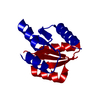
| Structure viewer | Molecule: MolmilJmol/JSmol |
|---|
- Downloads & links
-Download
| PDBx/mmCIF format | 1kzb.cif.gz | 65.5 KB | Display | PDBx/mmCIF format |
|---|---|---|---|---|
| PDB format | pdb1kzb.ent.gz | 46.7 KB | Display | PDB format |
| PDBx/mmJSON format | 1kzb.json.gz | Tree view | PDBx/mmJSON format | |
| Others |  Other downloads Other downloads |
-Validation report
| Arichive directory | https://data.pdbj.org/pub/pdb/validation_reports/kz/1kzbftp://data.pdbj.org/pub/pdb/validation_reports/kz/1kzb | HTTPS FTP |
|---|
-Related structure data
| Related structure data |  1kwtC  1kwuC  1kwvC 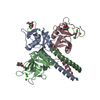 1kwwC  1kwxC 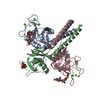 1kwyC 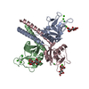 1kwzC 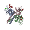 1kx0C  1kx1C  1kzaC 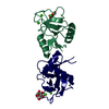 1kzcC 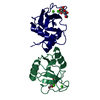 1kzdC  1kzeC 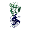 1rdoS S: Starting model for refinement C: citing same article ( |
|---|---|
| Similar structure data |


-Links
 PDBj
PDBj


- Assembly
| Deposited unit | 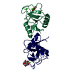
| ||||||||||
|---|---|---|---|---|---|---|---|---|---|---|---|
| 1 |
| ||||||||||
| Unit cell |
| ||||||||||
| Details | non-physiological dimer |
-Components
| #1: Protein | Mass: 12830.325 Da / Num. of mol.: 2 / Fragment: SUBTILISIN FRAGMENT (RESIDUES 129-243 of P08661) Source method: isolated from a genetically manipulated source Source: (gene. exp.)  #2: Sugar |   Type: D-saccharide, alpha linking / Mass: 180.156 Da / Num. of mol.: 2 Type: D-saccharide, alpha linking / Mass: 180.156 Da / Num. of mol.: 2Source method: isolated from a genetically manipulated source Formula: C6H12O6 #3: Chemical | ChemComp-CA /   Mass: 40.078 Da / Num. of mol.: 4 / Source method: obtained synthetically / Formula: Ca Mass: 40.078 Da / Num. of mol.: 4 / Source method: obtained synthetically / Formula: Ca#4: Water | ChemComp-HOH / |  Mass: 18.015 Da / Num. of mol.: 293 / Source method: isolated from a natural source / Formula: H2O Mass: 18.015 Da / Num. of mol.: 293 / Source method: isolated from a natural source / Formula: H2OHas protein modification | Y | |
|---|
-Experimental details
-Experiment
| Experiment | Method: X-RAY DIFFRACTION / Number of used crystals: 1 |
|---|
- Sample preparation
| Crystal | Density Matthews: 2.56 Å3/Da / Density % sol: 51.93 % | |||||||||||||||||||||||||||||||||||||||||||||||||||||||||||||||
|---|---|---|---|---|---|---|---|---|---|---|---|---|---|---|---|---|---|---|---|---|---|---|---|---|---|---|---|---|---|---|---|---|---|---|---|---|---|---|---|---|---|---|---|---|---|---|---|---|---|---|---|---|---|---|---|---|---|---|---|---|---|---|---|---|
| Crystal grow | Temperature: 298 K / Method: vapor diffusion, hanging drop / pH: 7.4 Details: PEG 8000, Tris-Cl, NaCl, CaCl2, NaN3, pH 7.4, VAPOR DIFFUSION, HANGING DROP at 298K | |||||||||||||||||||||||||||||||||||||||||||||||||||||||||||||||
| Crystal grow | *PLUS Temperature: 20-22 ℃ / pH: 7.5 | |||||||||||||||||||||||||||||||||||||||||||||||||||||||||||||||
| Components of the solutions | *PLUS
|
-Data collection
| Diffraction | Mean temperature: 100 K |
|---|---|
| Diffraction source | Source: ROTATING ANODE / Type: RIGAKU RU200 / Wavelength: 1.5418 Å |
| Detector | Type: RIGAKU RAXIS IIC / Detector: IMAGE PLATE / Date: Nov 10, 1994 |
| Radiation | Monochromator: graphite / Protocol: SINGLE WAVELENGTH / Monochromatic (M) / Laue (L): M / Scattering type: x-ray |
| Radiation wavelength | Wavelength: 1.5418 Å / Relative weight: 1 |
| Reflection | Resolution: 1.8→40 Å / Num. all: 23828 / Num. obs: 23828 / % possible obs: 94.8 % / Observed criterion σ(I): -3 / Redundancy: 3.3 % / Rmerge(I) obs: 0.068 / Rsym value: 0.068 / Net I/σ(I): 17.3 |
| Reflection shell | Resolution: 1.8→1.86 Å / Redundancy: 1.5 % / Rmerge(I) obs: 0.111 / Mean I/σ(I) obs: 4 / Num. unique all: 2006 / Rsym value: 0.111 / % possible all: 81.8 |
| Reflection | *PLUS Highest resolution: 1.8 Å / Lowest resolution: 40 Å / Redundancy: 3.5 % |
| Reflection shell | *PLUS % possible obs: 81.8 % / Redundancy: 1.8 % |
- Processing
| Software |
| |||||||||||||||||||||||||
|---|---|---|---|---|---|---|---|---|---|---|---|---|---|---|---|---|---|---|---|---|---|---|---|---|---|---|
| Refinement | Method to determine structure: FOURIER SYNTHESIS Starting model: PDB entry 1RDO Resolution: 1.8→40 Å / Isotropic thermal model: isotropic / Cross valid method: THROUGHOUT / σ(F): 0 / σ(I): 0 / Stereochemistry target values: Engh & Huber
| |||||||||||||||||||||||||
| Displacement parameters |
| |||||||||||||||||||||||||
| Refinement step | Cycle: LAST / Resolution: 1.8→40 Å
| |||||||||||||||||||||||||
| Refine LS restraints |
| |||||||||||||||||||||||||
| Refinement | *PLUS Highest resolution: 1.8 Å / Lowest resolution: 40 Å | |||||||||||||||||||||||||
| Solvent computation | *PLUS | |||||||||||||||||||||||||
| Displacement parameters | *PLUS | |||||||||||||||||||||||||
| Refine LS restraints | *PLUS Type: c_angle_d |
