 Movie
Movie Controller

[English] 日本語
 Yorodumi
- PDB-4ut5: Crystal structure of the LecB lectin from Pseudomonas aeruginosa ... -
+ Open data

- Basic information
| Entry | Database: PDB / ID: 4ut5 | |||||||||
|---|---|---|---|---|---|---|---|---|---|---|
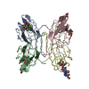
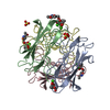
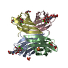
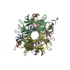
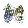
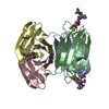
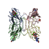
| Title | Crystal structure of the LecB lectin from Pseudomonas aeruginosa strain PA7 in complex with lewis a tetrasaccharide | |||||||||
 Components Components | LECB LECTIN | |||||||||
 Keywords Keywords | SUGAR BINDING PROTEIN / LECTIN / TETRAMERIC / LEWIS A / LECB VARIANT | |||||||||
| Function / homology |  Function and homology information Function and homology information | |||||||||
| Biological species |   PSEUDOMONAS AERUGINOSA (bacteria) PSEUDOMONAS AERUGINOSA (bacteria) | |||||||||
| Method |  X-RAY DIFFRACTION / SYNCHROTRON / MOLECULAR REPLACEMENT / Resolution: 1.75 Å X-RAY DIFFRACTION / SYNCHROTRON / MOLECULAR REPLACEMENT / Resolution: 1.75 Å | |||||||||
 Authors Authors | Boukerb, A.M. / Decor, A. / Tabaroni, R. / Varrot, A. / Debentzmann, S. / Vidal, S. / Imberty, A. / Cournoyer, B. | |||||||||
 Citation Citation | Journal: Front.Microbiol. / Year: 2016 Title: Genomic Rearrangements and Functional Diversification of Leca and Lecb Lectin-Coding Regions Impacting the Efficacy of Glycomimetics Directed Against Pseudomonas Aeruginosa. Authors: Boukerb, A.M. / Decor, A. / Ribun, S. / Tabaroni, R. / Rousset, A. / Commin, L. / Buff, S. / Doleans-Jordheim, A. / Vidal, S. / Varrot, A. / Imberty, A. / Cournoyer, B. | |||||||||
| History |
| |||||||||
| Remark 700 | SHEET DETERMINATION METHOD: AUTHOR PROVIDED. |
- Structure visualization
| Structure viewer | Molecule: MolmilJmol/JSmol |
|---|
- Downloads & links
-Download
| PDBx/mmCIF format | 4ut5.cif.gz | 117.4 KB | Display | PDBx/mmCIF format |
|---|---|---|---|---|
| PDB format | pdb4ut5.ent.gz | 90.8 KB | Display | PDB format |
| PDBx/mmJSON format | 4ut5.json.gz | Tree view | PDBx/mmJSON format | |
| Others |  Other downloads Other downloads |
-Validation report
| Arichive directory | https://data.pdbj.org/pub/pdb/validation_reports/ut/4ut5ftp://data.pdbj.org/pub/pdb/validation_reports/ut/4ut5 | HTTPS FTP |
|---|
-Related structure data
| Related structure data |  1gztS S: Starting model for refinement |
|---|---|
| Similar structure data |


-Links
 PDBj
PDBj

- Assembly
| Deposited unit | 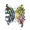
| ||||||||||||||||
|---|---|---|---|---|---|---|---|---|---|---|---|---|---|---|---|---|---|
| 1 |
| ||||||||||||||||
| Unit cell |
| ||||||||||||||||
| Noncrystallographic symmetry (NCS) | NCS oper:
|
-Components
-Protein , 1 types, 4 molecules ABCD
| #1: Protein | Mass: 11659.705 Da / Num. of mol.: 4 Source method: isolated from a genetically manipulated source Source: (gene. exp.) PSEUDOMONAS AERUGINOSA (bacteria) / Strain: PA7 / Plasmid: PFQ1044 / Production host: |
|---|
-Sugars , 4 types, 5 molecules 
| #2: Polysaccharide | beta-D-galactopyranose-(1-3)-[alpha-L-fucopyranose-(1-4)]2-acetamido-2-deoxy-beta-D-glucopyranose- ...beta-D-galactopyranose-(1-3)-[alpha-L-fucopyranose-(1-4)]2-acetamido-2-deoxy-beta-D-glucopyranose-(1-3)-beta-D-galactopyranose Source method: isolated from a genetically manipulated source | ||||
|---|---|---|---|---|---|
| #3: Polysaccharide | Source method: isolated from a genetically manipulated source #4: Polysaccharide | beta-D-galactopyranose-(1-3)-[alpha-L-fucopyranose-(1-4)]2-acetamido-2-deoxy-beta-D-glucopyranose | Source method: isolated from a genetically manipulated source #6: Sugar | ChemComp-GAL / |  Type: D-saccharide, beta linking / Mass: 180.156 Da / Num. of mol.: 1 Type: D-saccharide, beta linking / Mass: 180.156 Da / Num. of mol.: 1Source method: isolated from a genetically manipulated source Formula: C6H12O6 |
-Non-polymers , 2 types, 652 molecules 

| #5: Chemical | ChemComp-CA /  Mass: 40.078 Da / Num. of mol.: 8 / Source method: obtained synthetically / Formula: Ca Mass: 40.078 Da / Num. of mol.: 8 / Source method: obtained synthetically / Formula: Ca#7: Water | ChemComp-HOH / | Mass: 18.015 Da / Num. of mol.: 644 / Source method: isolated from a natural source / Formula: H2O |
|---|
-Experimental details
-Experiment
| Experiment | Method: X-RAY DIFFRACTION / Number of used crystals: 1 |
|---|
- Sample preparation
| Crystal | Density Matthews: 2.16 Å3/Da / Density % sol: 43.08 % / Description: NONE |
|---|---|
| Crystal grow | pH: 4.6 Details: 14% PEG6K, 0.2M LITHIUM CHLORIDE AND 0.1M SODIUM ACETATE PH 4.6. CRYO WITH 26% PEG 6K |
-Data collection
| Diffraction | Mean temperature: 100 K |
|---|---|
| Diffraction source | Source: SYNCHROTRON / Site: ESRF  / Beamline: BM30A / Wavelength: 0.9205 / Beamline: BM30A / Wavelength: 0.9205 |
| Detector | Type: MARRESEARCH / Detector: CCD / Date: Apr 7, 2014 |
| Radiation | Protocol: SINGLE WAVELENGTH / Monochromatic (M) / Laue (L): M / Scattering type: x-ray |
| Radiation wavelength | Wavelength: 0.9205 Å / Relative weight: 1 |
| Reflection | Resolution: 1.75→42.23 Å / Num. obs: 40169 / % possible obs: 99.4 % / Observed criterion σ(I): 2 / Redundancy: 3.4 % / Rmerge(I) obs: 0.06 / Net I/σ(I): 13.7 |
| Reflection shell | Resolution: 1.75→1.78 Å / Redundancy: 2.7 % / Rmerge(I) obs: 0.35 / Mean I/σ(I) obs: 3 / % possible all: 93.5 |
- Processing
| Software |
| ||||||||||||||||||||||||||||||||||||||||||||||||||||||||||||||||||||||||||||||||||||||||||||||||||||||||||||||||||||||||||||||||||||||||||||||||||||||||||||||||||||||||||||||||||||||
|---|---|---|---|---|---|---|---|---|---|---|---|---|---|---|---|---|---|---|---|---|---|---|---|---|---|---|---|---|---|---|---|---|---|---|---|---|---|---|---|---|---|---|---|---|---|---|---|---|---|---|---|---|---|---|---|---|---|---|---|---|---|---|---|---|---|---|---|---|---|---|---|---|---|---|---|---|---|---|---|---|---|---|---|---|---|---|---|---|---|---|---|---|---|---|---|---|---|---|---|---|---|---|---|---|---|---|---|---|---|---|---|---|---|---|---|---|---|---|---|---|---|---|---|---|---|---|---|---|---|---|---|---|---|---|---|---|---|---|---|---|---|---|---|---|---|---|---|---|---|---|---|---|---|---|---|---|---|---|---|---|---|---|---|---|---|---|---|---|---|---|---|---|---|---|---|---|---|---|---|---|---|---|---|
| Refinement | Method to determine structure: MOLECULAR REPLACEMENT Starting model: PDB ENTRY 1GZT Resolution: 1.75→42.23 Å / Cor.coef. Fo:Fc: 0.96 / Cor.coef. Fo:Fc free: 0.949 / SU B: 2.178 / SU ML: 0.069 / Cross valid method: THROUGHOUT / ESU R: 0.116 / ESU R Free: 0.108 / Stereochemistry target values: MAXIMUM LIKELIHOOD Details: HYDROGENS HAVE BEEN ADDED IN THE RIDING POSITIONS. U VALUES REFINED INDIVIDUALLY DISORDERED REGIONS WERE MODELED STEREOCHEMICALLY THE NUMBERING IS SHEFTED BY ONE COMPARE TO THE UNIPROT ...Details: HYDROGENS HAVE BEEN ADDED IN THE RIDING POSITIONS. U VALUES REFINED INDIVIDUALLY DISORDERED REGIONS WERE MODELED STEREOCHEMICALLY THE NUMBERING IS SHEFTED BY ONE COMPARE TO THE UNIPROT SEQUENCE SINCE THE NTERMINAL METHIONINE IS NOT PRESENT IN THE MATURE PROTEIN
| ||||||||||||||||||||||||||||||||||||||||||||||||||||||||||||||||||||||||||||||||||||||||||||||||||||||||||||||||||||||||||||||||||||||||||||||||||||||||||||||||||||||||||||||||||||||
| Solvent computation | Ion probe radii: 0.8 Å / Shrinkage radii: 0.8 Å / VDW probe radii: 1.2 Å / Solvent model: MASK | ||||||||||||||||||||||||||||||||||||||||||||||||||||||||||||||||||||||||||||||||||||||||||||||||||||||||||||||||||||||||||||||||||||||||||||||||||||||||||||||||||||||||||||||||||||||
| Displacement parameters | Biso mean: 12.49 Å2
| ||||||||||||||||||||||||||||||||||||||||||||||||||||||||||||||||||||||||||||||||||||||||||||||||||||||||||||||||||||||||||||||||||||||||||||||||||||||||||||||||||||||||||||||||||||||
| Refinement step | Cycle: LAST / Resolution: 1.75→42.23 Å
| ||||||||||||||||||||||||||||||||||||||||||||||||||||||||||||||||||||||||||||||||||||||||||||||||||||||||||||||||||||||||||||||||||||||||||||||||||||||||||||||||||||||||||||||||||||||
| Refine LS restraints |
|