 Movie
Movie Controller

[English] 日本語
 Yorodumi
- PDB-5nes: Discovery, crystal structures and atomic force microscopy study o... -
+ Open data

- Basic information
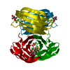
| Entry | Database: PDB / ID: 5nes | ||||||
|---|---|---|---|---|---|---|---|
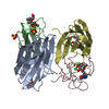
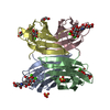
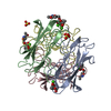
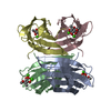
| Title | Discovery, crystal structures and atomic force microscopy study of thioether ligated D,L-cyclic antimicrobial peptides against multidrug resistant Pseudomonas aeruginosa | ||||||
 Components Components |
| ||||||
 Keywords Keywords | SUGAR BINDING PROTEIN / Cyclic peptides / Antimicrobials / Pseudomonas aeruginosa | ||||||
| Function / homology |  Function and homology information Function and homology informationsingle-species biofilm formation / carbohydrate binding / metal ion binding Similarity search - Function | ||||||
| Biological species |   Pseudomonas aeruginosa (bacteria) Pseudomonas aeruginosa (bacteria)synthetic construct (others) | ||||||
| Method |  X-RAY DIFFRACTION / SYNCHROTRON / MOLECULAR REPLACEMENT / Resolution: 1.606 Å X-RAY DIFFRACTION / SYNCHROTRON / MOLECULAR REPLACEMENT / Resolution: 1.606 Å | ||||||
 Authors Authors | Reymond, J.-L. / Darbre, T. / Stocker, A. / Hong, W. / van Delden, C. / Koehler, T. / Luscher, A. / Visini, R. / Fu, Y. / Di Bonaventura, I. / He, R. | ||||||
 Citation Citation | Journal: Chem Sci / Year: 2017 Title: Design, crystal structure and atomic force microscopy study of thioether ligated d,l-cyclic antimicrobial peptides against multidrug resistant Pseudomonas aeruginosa. Authors: He, R. / Di Bonaventura, I. / Visini, R. / Gan, B.H. / Fu, Y. / Probst, D. / Luscher, A. / Kohler, T. / van Delden, C. / Stocker, A. / Hong, W. / Darbre, T. / Reymond, J.L. | ||||||
| History |
|
- Structure visualization
| Structure viewer | Molecule: MolmilJmol/JSmol |
|---|
- Downloads & links
-Download
| PDBx/mmCIF format | 5nes.cif.gz | 115.6 KB | Display | PDBx/mmCIF format |
|---|---|---|---|---|
| PDB format | pdb5nes.ent.gz | 87.5 KB | Display | PDB format |
| PDBx/mmJSON format | 5nes.json.gz | Tree view | PDBx/mmJSON format | |
| Others |  Other downloads Other downloads |
-Validation report
| Arichive directory | https://data.pdbj.org/pub/pdb/validation_reports/ne/5nesftp://data.pdbj.org/pub/pdb/validation_reports/ne/5nes | HTTPS FTP |
|---|
-Related structure data
| Related structure data |  5neyC  5nf0C  1oxcS S: Starting model for refinement C: citing same article ( |
|---|---|
| Similar structure data |


-Links
 PDBj
PDBj- Assembly
| Deposited unit | 
| ||||||||
|---|---|---|---|---|---|---|---|---|---|
| 1 |
| ||||||||
| Unit cell |
|
-Components
-Protein / Protein/peptide / Sugars , 3 types, 9 molecules ABCDE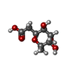
| #1: Protein | Mass: 11734.707 Da / Num. of mol.: 4 Source method: isolated from a genetically manipulated source Source: (gene. exp.) Pseudomonas aeruginosa (bacteria)Gene: lecB, PAERUG_E15_London_28_01_14_00983, PAERUG_P32_London_17_VIM_2_10_11_00423, PAMH19_1713 Production host: #2: Protein/peptide | | Mass: 1613.025 Da / Num. of mol.: 1 / Source method: obtained synthetically / Source: (synth.) synthetic construct (others) #4: Sugar | ChemComp-ZDC /  Type: D-saccharide / Mass: 206.193 Da / Num. of mol.: 4 Type: D-saccharide / Mass: 206.193 Da / Num. of mol.: 4Source method: isolated from a genetically manipulated source Formula: C8H14O6 |
|---|
-Non-polymers , 3 types, 581 molecules 
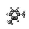

| #3: Chemical | ChemComp-CA /  Mass: 40.078 Da / Num. of mol.: 8 / Source method: obtained synthetically / Formula: Ca Mass: 40.078 Da / Num. of mol.: 8 / Source method: obtained synthetically / Formula: Ca#5: Chemical | ChemComp-8VH / |  Mass: 106.165 Da / Num. of mol.: 1 Mass: 106.165 Da / Num. of mol.: 1Source method: isolated from a genetically manipulated source Formula: C8H10 #6: Water | ChemComp-HOH / | Mass: 18.015 Da / Num. of mol.: 572 / Source method: isolated from a natural source / Formula: H2O |
|---|
-Details
| Has protein modification | Y |
|---|
-Experimental details
-Experiment
| Experiment | Method: X-RAY DIFFRACTION / Number of used crystals: 1 |
|---|
- Sample preparation
| Crystal | Density Matthews: 2.27 Å3/Da / Density % sol: 45.76 % |
|---|---|
| Crystal grow | Temperature: 291 K / Method: vapor diffusion, sitting drop / pH: 7.5 Details: 0.005 M Cobalt(II) chloride hexahydrate, 0.1M HEPES, 12% Polyethylene glycol 3350, |
-Data collection
| Diffraction | Mean temperature: 100 K |
|---|---|
| Diffraction source | Source: SYNCHROTRON / Site: SLS  / Beamline: X06DA / Wavelength: 1.03679 Å / Beamline: X06DA / Wavelength: 1.03679 Å |
| Detector | Type: DECTRIS PILATUS 2M / Detector: PIXEL / Date: Aug 20, 2016 |
| Radiation | Protocol: MAD / Monochromatic (M) / Laue (L): M / Scattering type: x-ray |
| Radiation wavelength | Wavelength: 1.03679 Å / Relative weight: 1 |
| Reflection | Resolution: 1.606→47.784 Å / Num. obs: 51247 / % possible obs: 90.6 % / Redundancy: 1.78 % / Rrim(I) all: 0.032 / Net I/σ(I): 23.48 |
| Reflection shell | Resolution: 1.606→1.7 Å / Mean I/σ(I) obs: 11.32 / Num. unique all: 7617 / CC1/2: 0.99 / % possible all: 82.9 |
- Processing
| Software |
| |||||||||||||||||||||||||||||||||||||||||||||||||||||||||||||||||||||||||||||||||||||||||||||||||||||||||||||||||||||||||||||||||||||
|---|---|---|---|---|---|---|---|---|---|---|---|---|---|---|---|---|---|---|---|---|---|---|---|---|---|---|---|---|---|---|---|---|---|---|---|---|---|---|---|---|---|---|---|---|---|---|---|---|---|---|---|---|---|---|---|---|---|---|---|---|---|---|---|---|---|---|---|---|---|---|---|---|---|---|---|---|---|---|---|---|---|---|---|---|---|---|---|---|---|---|---|---|---|---|---|---|---|---|---|---|---|---|---|---|---|---|---|---|---|---|---|---|---|---|---|---|---|---|---|---|---|---|---|---|---|---|---|---|---|---|---|---|---|---|
| Refinement | Method to determine structure: MOLECULAR REPLACEMENT Starting model: 1OXC Resolution: 1.606→47.784 Å / SU ML: 0.13 / Cross valid method: FREE R-VALUE / σ(F): 2.02 / Phase error: 15.61
| |||||||||||||||||||||||||||||||||||||||||||||||||||||||||||||||||||||||||||||||||||||||||||||||||||||||||||||||||||||||||||||||||||||
| Solvent computation | Shrinkage radii: 0.9 Å / VDW probe radii: 1.11 Å | |||||||||||||||||||||||||||||||||||||||||||||||||||||||||||||||||||||||||||||||||||||||||||||||||||||||||||||||||||||||||||||||||||||
| Refinement step | Cycle: LAST / Resolution: 1.606→47.784 Å
| |||||||||||||||||||||||||||||||||||||||||||||||||||||||||||||||||||||||||||||||||||||||||||||||||||||||||||||||||||||||||||||||||||||
| Refine LS restraints |
| |||||||||||||||||||||||||||||||||||||||||||||||||||||||||||||||||||||||||||||||||||||||||||||||||||||||||||||||||||||||||||||||||||||
| LS refinement shell |
|
