Mass: 18.015 Da / Num. of mol.: 22 / Source method: isolated from a natural source / Formula: H2O
-
Details
Nonpolymer details
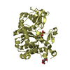
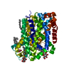
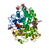
L-TRYPTOPHAN (TRP): SUBSTRATE
Sequence details
THE LEADING THREE RESIDUES ARE LEFTOVER FROM THE EXPRESSION CONSTRUCT WITH THE LEAD METHIONINE ...THE LEADING THREE RESIDUES ARE LEFTOVER FROM THE EXPRESSION CONSTRUCT WITH THE LEAD METHIONINE REMOVED. NUMBERING IN THE PDB FILE REFLECTS THE UNIPROT SEQUENCE AND NUMBERING
-
Experimental details
-
Experiment
Experiment
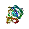
Method: X-RAY DIFFRACTION / Number of used crystals: 1
-
Sample preparation
Crystal
Density Matthews: 2.37 Å3/Da / Density % sol: 48 % / Description: NONE
Crystal grow
Method: lipidic cubic phase / pH: 7 Details: CRYSTALLISED BY LCP USING 7.8 MAG AS HOST LIPID. CRYSTALS WERE GROWN IN 20% PEG400, 400 MM NACL, 100 MM HEPES PH 7.0
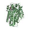
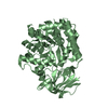
Resolution: 2.6→39.442 Å / SU ML: 0.43 / σ(F): 1.34 / Phase error: 26.95 / Stereochemistry target values: ML Details: RESIDUES 17-449 BUILT IN THE STRUCTURE. RESIDUE 449 BUILT ONLY AS BACKBONE AS SIDE CHAIN IS DISORDERED
Rfactor
Num. reflection
% reflection
Rfree
0.2545
1003
7.5 %
Rwork
0.201
-
-
obs
0.205
13303
96.31 %
Solvent computation
Shrinkage radii: 0.9 Å / VDW probe radii: 1.11 Å / Solvent model: FLAT BULK SOLVENT MODEL
Displacement parameters
Biso mean: 46.2 Å2
Refinement step
Cycle: LAST / Resolution: 2.6→39.442 Å
Protein
Nucleic acid
Ligand
Solvent
Total
Num. atoms
3243
0
83
22
3348
Refine LS restraints
Refine-ID
Type
Dev ideal
Number
X-RAY DIFFRACTION
f_bond_d
0.008
3423
X-RAY DIFFRACTION
f_angle_d
0.81
4658
X-RAY DIFFRACTION
f_dihedral_angle_d
11.417
1169
X-RAY DIFFRACTION
f_chiral_restr
0.026
536
X-RAY DIFFRACTION
f_plane_restr
0.005
561
LS refinement shell
Resolution (Å)
Rfactor Rfree
Num. reflection Rfree
Rfactor Rwork
Num. reflection Rwork
Refine-ID
% reflection obs (%)
2.6001-2.7371
0.3832
143
0.2834
1749
X-RAY DIFFRACTION
97
2.7371-2.9086
0.3239
140
0.2578
1752
X-RAY DIFFRACTION
96
2.9086-3.1331
0.3155
144
0.2359
1740
X-RAY DIFFRACTION
96
3.1331-3.4482
0.2985
143
0.2089
1768
X-RAY DIFFRACTION
97
3.4482-3.9467
0.2405
146
0.177
1743
X-RAY DIFFRACTION
96
3.9467-4.9709
0.2018
141
0.1719
1761
X-RAY DIFFRACTION
97
4.9709-39.4467
0.216
146
0.1879
1787
X-RAY DIFFRACTION
96
+
About Yorodumi
-
News
-
Feb 9, 2022. New format data for meta-information of EMDB entries
New format data for meta-information of EMDB entries
Version 3 of the EMDB header file is now the official format.
The previous official version 1.9 will be removed from the archive.
In the structure databanks used in Yorodumi, some data are registered as the other names, "COVID-19 virus" and "2019-nCoV". Here are the details of the virus and the list of structure data.
Jan 31, 2019. EMDB accession codes are about to change! (news from PDBe EMDB page)
EMDB accession codes are about to change! (news from PDBe EMDB page)
The allocation of 4 digits for EMDB accession codes will soon come to an end. Whilst these codes will remain in use, new EMDB accession codes will include an additional digit and will expand incrementally as the available range of codes is exhausted. The current 4-digit format prefixed with “EMD-” (i.e. EMD-XXXX) will advance to a 5-digit format (i.e. EMD-XXXXX), and so on. It is currently estimated that the 4-digit codes will be depleted around Spring 2019, at which point the 5-digit format will come into force.
The EM Navigator/Yorodumi systems omit the EMD- prefix.
Related info.:Q: What is EMD? / ID/Accession-code notation in Yorodumi/EM Navigator
Yorodumi is a browser for structure data from EMDB, PDB, SASBDB, etc.
This page is also the successor to EM Navigator detail page, and also detail information page/front-end page for Omokage search.
The word "yorodu" (or yorozu) is an old Japanese word meaning "ten thousand". "mi" (miru) is to see.
Related info.:EMDB / PDB / SASBDB / Comparison of 3 databanks / Yorodumi Search / Aug 31, 2016. New EM Navigator & Yorodumi / Yorodumi Papers / Jmol/JSmol / Function and homology information / Changes in new EM Navigator and Yorodumi
 Movie
Movie Controller

 Yorodumi
 Open data
 Basic information
 Components
 Keywords
 Function and homology information
 BACILLUS HALODURANS (bacteria)
 X-RAY DIFFRACTION /
  Authors
 Citation
 Structure visualization
 Downloads & links
 Other downloads


 PDBj
PDBj


 Assembly
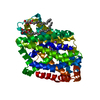


 Type: L-peptide linking / Mass: 204.225 Da / Num. of mol.: 1 / Source method: obtained synthetically / Formula: C11H12N2O2
 Mass: 22.990 Da / Num. of mol.: 2 / Source method: obtained synthetically / Formula: Na
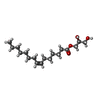
 Mass: 314.460 Da / Num. of mol.: 2 / Source method: obtained synthetically / Formula: C18H34O4
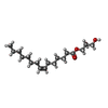
 Mass: 314.460 Da / Num. of mol.: 1 / Source method: obtained synthetically / Formula: C18H34O4
 Sample preparation
 / Beamline: I24 / Wavelength: 0.9686
  Processing