 Movie
Movie Controller

[English] 日本語
 Yorodumi
- PDB-3hpa: Crystal structure of an amidohydrolase gi:44264246 from an eviron... -
+ Open data

- Basic information
| Entry | Database: PDB / ID: 3hpa | |||||||||
|---|---|---|---|---|---|---|---|---|---|---|
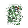
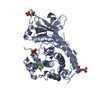
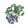
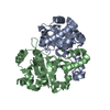
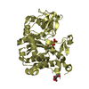
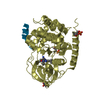
| Title | Crystal structure of an amidohydrolase gi:44264246 from an evironmental sample of sargasso sea | |||||||||
 Components Components | AMIDOHYDROLASE | |||||||||
 Keywords Keywords | HYDROLASE / AMIDOHYDROLASE / SIGNATURE OF ZN LIGANDS / STRUCTURAL GENOMICS / NYSGXRC / TARGET 9236E / PSI-2 / PROTEIN STRUCTURE INITIATIVE / NEW YORK SGX RESEARCH CENTER FOR STRUCTURAL GENOMICS | |||||||||
| Function / homology | Urease, subunit C; domain 1 / Urease, subunit C, domain 1 / Metal-dependent hydrolases / Roll / TIM Barrel / Alpha-Beta Barrel / Mainly Beta / Alpha Beta Function and homology information Function and homology information | |||||||||
| Biological species | unidentified (others) | |||||||||
| Method |  X-RAY DIFFRACTION / SYNCHROTRON / MOLECULAR REPLACEMENT / Resolution: 2.2 Å X-RAY DIFFRACTION / SYNCHROTRON / MOLECULAR REPLACEMENT / Resolution: 2.2 Å | |||||||||
 Authors Authors | Fedorov, A.A. / Fedorov, E.V. / Toro, R. / Raushel, F.M. / Burley, S.K. / Almo, S.C. / New York SGX Research Center for Structural Genomics (NYSGXRC) | |||||||||
 Citation Citation | Journal: J.Am.Chem.Soc. / Year: 2010 Title: The hunt for 8-oxoguanine deaminase. Authors: Hall, R.S. / Fedorov, A.A. / Marti-Arbona, R. / Fedorov, E.V. / Kolb, P. / Sauder, J.M. / Burley, S.K. / Shoichet, B.K. / Almo, S.C. / Raushel, F.M. | |||||||||
| History |
|
- Structure visualization
| Structure viewer | Molecule: MolmilJmol/JSmol |
|---|
- Downloads & links
-Download
| PDBx/mmCIF format | 3hpa.cif.gz | 172.1 KB | Display | PDBx/mmCIF format |
|---|---|---|---|---|
| PDB format | pdb3hpa.ent.gz | 135.9 KB | Display | PDB format |
| PDBx/mmJSON format | 3hpa.json.gz | Tree view | PDBx/mmJSON format | |
| Others |  Other downloads Other downloads |
-Validation report
| Arichive directory | https://data.pdbj.org/pub/pdb/validation_reports/hp/3hpaftp://data.pdbj.org/pub/pdb/validation_reports/hp/3hpa | HTTPS FTP |
|---|
-Related structure data
| Similar structure data | |
|---|---|
| Other databases |


-Links
 PDBj
PDBj- Assembly
| Deposited unit | 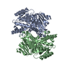
| ||||||||
|---|---|---|---|---|---|---|---|---|---|
| 1 | 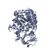
| ||||||||
| 2 | 
| ||||||||
| 3 |
| ||||||||
| Unit cell |
| ||||||||
| Components on special symmetry positions |
|
-Components
| #1: Protein | Mass: 51835.836 Da / Num. of mol.: 2 Source method: isolated from a genetically manipulated source Details: EVIRONMENTAL SAMPLE OF SARGASSO SEA / Source: (gene. exp.) unidentified (others) / Production host:  #2: Chemical |   Mass: 65.409 Da / Num. of mol.: 2 / Source method: obtained synthetically / Formula: Zn Mass: 65.409 Da / Num. of mol.: 2 / Source method: obtained synthetically / Formula: Zn#3: Water | ChemComp-HOH / |  Mass: 18.015 Da / Num. of mol.: 129 / Source method: isolated from a natural source / Formula: H2O Mass: 18.015 Da / Num. of mol.: 129 / Source method: isolated from a natural source / Formula: H2O |
|---|
-Experimental details
-Experiment
| Experiment | Method: X-RAY DIFFRACTION / Number of used crystals: 1 |
|---|
- Sample preparation
| Crystal | Density Matthews: 2.21 Å3/Da / Density % sol: 44.44 % |
|---|---|
| Crystal grow | Temperature: 293 K / Method: vapor diffusion, hanging drop / pH: 7 Details: 2.8M SODIUM ACETATE, pH 7.0, VAPOR DIFFUSION, HANGING DROP, temperature 293.0K |
-Data collection
| Diffraction | Mean temperature: 100 K |
|---|---|
| Diffraction source | Source: SYNCHROTRON / Site: NSLS  / Beamline: X4A / Wavelength: 0.97915 Å / Beamline: X4A / Wavelength: 0.97915 Å |
| Detector | Type: ADSC QUANTUM 4 / Detector: CCD / Date: Aug 15, 2008 |
| Radiation | Monochromator: Si 111 CHANNEL / Protocol: SINGLE WAVELENGTH / Monochromatic (M) / Laue (L): M / Scattering type: x-ray |
| Radiation wavelength | Wavelength: 0.97915 Å / Relative weight: 1 |
| Reflection | Resolution: 2.2→25 Å / Num. all: 47567 / Num. obs: 47567 / % possible obs: 99.7 % / Observed criterion σ(F): 0 / Observed criterion σ(I): 0 / Biso Wilson estimate: 27.9 Å2 / Rmerge(I) obs: 0.079 |
- Processing
| Software |
| ||||||||||||||||||||||||||||||||||||
|---|---|---|---|---|---|---|---|---|---|---|---|---|---|---|---|---|---|---|---|---|---|---|---|---|---|---|---|---|---|---|---|---|---|---|---|---|---|
| Refinement | Method to determine structure: MOLECULAR REPLACEMENT / Resolution: 2.2→24.62 Å / Rfactor Rfree error: 0.006 / Data cutoff high absF: 2073974.63 / Data cutoff low absF: 0 / Isotropic thermal model: RESTRAINED / Cross valid method: THROUGHOUT / σ(F): 0 / σ(I): 0 / Stereochemistry target values: Engh & Huber
| ||||||||||||||||||||||||||||||||||||
| Solvent computation | Solvent model: FLAT MODEL / Bsol: 62.4521 Å2 / ksol: 0.39385 e/Å3 | ||||||||||||||||||||||||||||||||||||
| Displacement parameters | Biso mean: 43.8 Å2
| ||||||||||||||||||||||||||||||||||||
| Refine analyze |
| ||||||||||||||||||||||||||||||||||||
| Refinement step | Cycle: LAST / Resolution: 2.2→24.62 Å
| ||||||||||||||||||||||||||||||||||||
| Refine LS restraints |
| ||||||||||||||||||||||||||||||||||||
| LS refinement shell | Resolution: 2.2→2.28 Å / Rfactor Rfree error: 0.021 / Total num. of bins used: 10
| ||||||||||||||||||||||||||||||||||||
| Xplor file |
|
