 Movie
Movie Controller


+ Open data

- Basic information
| Entry | Database: PDB / ID: 1pvw | ||||||
|---|---|---|---|---|---|---|---|
| Title | 3,4-dihydroxy-2-butanone 4-phosphate synthase from M. jannaschii | ||||||
 Components Components | 3,4-dihydroxy-2-butanone 4-phosphate synthase | ||||||
 Keywords Keywords | ISOMERASE / riboflavin biosynthesis | ||||||
| Function / homology |  Function and homology information Function and homology information3,4-dihydroxy-2-butanone-4-phosphate synthase / 3,4-dihydroxy-2-butanone-4-phosphate synthase activity / riboflavin biosynthetic process / manganese ion binding / magnesium ion binding / cytosol Similarity search - Function | ||||||
| Biological species |   Methanocaldococcus jannaschii (archaea) Methanocaldococcus jannaschii (archaea) | ||||||
| Method |  X-RAY DIFFRACTION / MIR / Resolution: 2.45 Å X-RAY DIFFRACTION / MIR / Resolution: 2.45 Å | ||||||
 Authors Authors | Steinbacher, S. / Schiffmann, S. / Richter, G. / Huber, R. / Bacher, A. / Fischer, M. | ||||||
 Citation Citation | Journal: J.Biol.Chem. / Year: 2003 Title: Structure of 3,4-Dihydroxy-2-butanone 4-Phosphate Synthase from Methanococcus jannaschii in Complex with Divalent Metal Ions and the Substrate Ribulose 5-Phosphate: IMPLICATIONS FOR THE CATALYTIC MECHANISM Authors: Steinbacher, S. / Schiffmann, S. / Richter, G. / Huber, R. / Bacher, A. / Fischer, M. #1: Journal: J.Biol.Chem. / Year: 2002Title: Biosynthesis of Riboflavin in Archaea Studies on the Mechanism of 3,4-Dihydroxy-2-butanone-4-phosphate Synthase of Methanococcus jannaschii Authors: Fischer, M. / Romisch, W. / Schiffmann, S. / Kelly, M. / Oschkinat, H. / Steinbacher, S. / Huber, R. / Eisenreich, W. / Richter, G. / Bacher, A. | ||||||
| History |
|
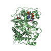
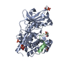
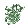
- Structure visualization
| Structure viewer | Molecule: MolmilJmol/JSmol |
|---|
- Downloads & links
-Download
| PDBx/mmCIF format | 1pvw.cif.gz | 138.8 KB | Display | PDBx/mmCIF format |
|---|---|---|---|---|
| PDB format | pdb1pvw.ent.gz | 110 KB | Display | PDB format |
| PDBx/mmJSON format | 1pvw.json.gz | Tree view | PDBx/mmJSON format | |
| Others |  Other downloads Other downloads |
-Validation report
| Arichive directory | https://data.pdbj.org/pub/pdb/validation_reports/pv/1pvwftp://data.pdbj.org/pub/pdb/validation_reports/pv/1pvw | HTTPS FTP |
|---|
-Related structure data


-Links
 PDBj
PDBj- Assembly
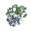
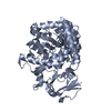
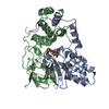
| Deposited unit | 
| ||||||||
|---|---|---|---|---|---|---|---|---|---|
| 1 | 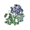
| ||||||||
| 2 | 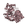
| ||||||||
| 3 | 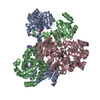
| ||||||||
| Unit cell |
| ||||||||
| Details | dimer; the asymmeric unit contains a dimer, representing the biological assembly and a monomer fro which the second monomer is generated by a twofold axis |
-Components
| #1: Protein | Mass: 25832.621 Da / Num. of mol.: 3 Source method: isolated from a genetically manipulated source Source: (gene. exp.) Methanocaldococcus jannaschii (archaea)Gene: MJ0055 / Plasmid: pNCO-MJ / Production host:  References: UniProt: Q60364, Isomerases; Intramolecular transferases; Transferring other groups #2: Chemical |   Mass: 65.409 Da / Num. of mol.: 2 / Source method: obtained synthetically / Formula: Zn Mass: 65.409 Da / Num. of mol.: 2 / Source method: obtained synthetically / Formula: Zn#3: Chemical | ChemComp-CA / |   Mass: 40.078 Da / Num. of mol.: 1 / Source method: obtained synthetically / Formula: Ca Mass: 40.078 Da / Num. of mol.: 1 / Source method: obtained synthetically / Formula: Ca#4: Chemical | ChemComp-PO4 / |   Mass: 94.971 Da / Num. of mol.: 1 / Source method: obtained synthetically / Formula: PO4 Mass: 94.971 Da / Num. of mol.: 1 / Source method: obtained synthetically / Formula: PO4#5: Water | ChemComp-HOH / |  Mass: 18.015 Da / Num. of mol.: 6 / Source method: isolated from a natural source / Formula: H2O Mass: 18.015 Da / Num. of mol.: 6 / Source method: isolated from a natural source / Formula: H2O |
|---|
-Experimental details
-Experiment
| Experiment | Method: X-RAY DIFFRACTION / Number of used crystals: 1 |
|---|
- Sample preparation
| Crystal | Density Matthews: 2.32 Å3/Da / Density % sol: 46.66 % |
|---|---|
| Crystal grow | Temperature: 293 K / Method: vapor diffusion, sitting drop / pH: 7.5 Details: PEG1000, pH 7.5, VAPOR DIFFUSION, SITTING DROP, temperature 293K |
| Crystal grow | *PLUS Method: unknown / Details: Fischer, M., (2002) J. Biol. Chem., 277, 41410. |
-Data collection
| Diffraction | Mean temperature: 287 K |
|---|---|
| Diffraction source | Source: ROTATING ANODE / Type: RIGAKU RU200 / Wavelength: 1.548 Å |
| Detector | Type: MARRESEARCH / Detector: IMAGE PLATE / Date: Jan 12, 2001 / Details: Osmic |
| Radiation | Protocol: SINGLE WAVELENGTH / Monochromatic (M) / Laue (L): M / Scattering type: x-ray |
| Radiation wavelength | Wavelength: 1.548 Å / Relative weight: 1 |
| Reflection | Resolution: 2.45→20 Å / Num. all: 24943 / Num. obs: 24943 / % possible obs: 97.8 % / Observed criterion σ(F): 0 / Observed criterion σ(I): 0 / Redundancy: 4.7 % / Rmerge(I) obs: 0.065 / Net I/σ(I): 21.5 |
| Reflection shell | Resolution: 2.45→2.54 Å / Rmerge(I) obs: 0.295 / Mean I/σ(I) obs: 3.5 / % possible all: 97.3 |
| Reflection shell | *PLUS % possible obs: 97.3 % |
- Processing
| Software |
| |||||||||||||||||||||||||
|---|---|---|---|---|---|---|---|---|---|---|---|---|---|---|---|---|---|---|---|---|---|---|---|---|---|---|
| Refinement | Method to determine structure: MIR / Resolution: 2.45→20 Å / Cross valid method: THROUGHOUT / σ(F): 0 / σ(I): 0 / Stereochemistry target values: Engh & Huber
| |||||||||||||||||||||||||
| Refinement step | Cycle: LAST / Resolution: 2.45→20 Å
| |||||||||||||||||||||||||
| Refine LS restraints |
| |||||||||||||||||||||||||
| Refinement | *PLUS Lowest resolution: 20 Å | |||||||||||||||||||||||||
| Solvent computation | *PLUS | |||||||||||||||||||||||||
| Displacement parameters | *PLUS |
