 Movie
Movie Controller

[English] 日本語
 Yorodumi
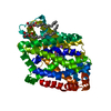
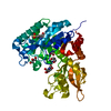
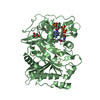
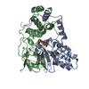
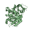
- PDB-3otk: Structure and mechanisim of core 2 beta1,6-n-acetylglucosaminyltr... -
+ Open data

- Basic information
| Entry | Database: PDB / ID: 3otk | |||||||||
|---|---|---|---|---|---|---|---|---|---|---|
| Title | Structure and mechanisim of core 2 beta1,6-n-acetylglucosaminyltransferase: a Metal-ion independent gt-a glycosyltransferase | |||||||||
 Components Components | Beta-1,3-galactosyl-O-glycosyl-glycoprotein beta-1,6-N-acetylglucosaminyltransferase | |||||||||
 Keywords Keywords | TRANSFERASE / glycosyltransferase / Golgi | |||||||||
| Function / homology |  Function and homology information Function and homology informationbeta-1,3-galactosyl-O-glycosyl-glycoprotein beta-1,6-N-acetylglucosaminyltransferase / beta-1,3-galactosyl-O-glycosyl-glycoprotein beta-1,6-N-acetylglucosaminyltransferase activity / O-linked glycosylation of mucins / tissue morphogenesis / kidney morphogenesis / protein O-linked glycosylation via N-acetylgalactosamine / Golgi cisterna / positive regulation of leukocyte tethering or rolling / cell adhesion molecule production / glycoprotein biosynthetic process ...beta-1,3-galactosyl-O-glycosyl-glycoprotein beta-1,6-N-acetylglucosaminyltransferase / beta-1,3-galactosyl-O-glycosyl-glycoprotein beta-1,6-N-acetylglucosaminyltransferase activity / O-linked glycosylation of mucins / tissue morphogenesis / kidney morphogenesis / protein O-linked glycosylation via N-acetylgalactosamine / Golgi cisterna / positive regulation of leukocyte tethering or rolling / cell adhesion molecule production / glycoprotein biosynthetic process / leukocyte tethering or rolling / trans-Golgi network / response to insulin / Golgi membrane / nucleotide binding / : Similarity search - Function | |||||||||
| Biological species |  | |||||||||
| Method |  X-RAY DIFFRACTION / SYNCHROTRON / MOLECULAR REPLACEMENT / Resolution: 2.3 Å X-RAY DIFFRACTION / SYNCHROTRON / MOLECULAR REPLACEMENT / Resolution: 2.3 Å | |||||||||
 Authors Authors | Pak, J.E. / Rini, J.M. | |||||||||
 Citation Citation | Journal: J.Mol.Biol. / Year: 2011 Title: Structural and mechanistic characterization of leukocyte-type core 2 beta 1,6-N-acetylglucosaminyltransferase: a metal-ion-independent GT-A glycosyltransferase. Authors: Pak, J.E. / Satkunarajah, M. / Seetharaman, J. / Rini, J.M. | |||||||||
| History |
|
- Structure visualization
| Structure viewer | Molecule: MolmilJmol/JSmol |
|---|
- Downloads & links
-Download
| PDBx/mmCIF format | 3otk.cif.gz | 587.8 KB | Display | PDBx/mmCIF format |
|---|---|---|---|---|
| PDB format | pdb3otk.ent.gz | 482.8 KB | Display | PDB format |
| PDBx/mmJSON format | 3otk.json.gz | Tree view | PDBx/mmJSON format | |
| Others |  Other downloads Other downloads |
-Validation report
| Arichive directory | https://data.pdbj.org/pub/pdb/validation_reports/ot/3otkftp://data.pdbj.org/pub/pdb/validation_reports/ot/3otk | HTTPS FTP |
|---|
-Related structure data
| Similar structure data |
|---|


-Links
 PDBj
PDBj


- Assembly
| Deposited unit | 
| ||||||||
|---|---|---|---|---|---|---|---|---|---|
| 1 | 
| ||||||||
| 2 | 
| ||||||||
| 3 | 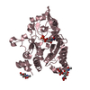
| ||||||||
| 4 | 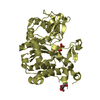
| ||||||||
| 5 | 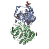
| ||||||||
| 6 | 
| ||||||||
| Unit cell |
|
-Components
-Protein , 1 types, 4 molecules ABCD
| #1: Protein | Mass: 45303.867 Da / Num. of mol.: 4 / Fragment: soluble catalytic domain (unp residues 38-428) / Mutation: C217S Source method: isolated from a genetically manipulated source Source: (gene. exp.) Cell (production host): HUMAN EMBRYONIC KIDNEY (HEK) 293 CELLS Production host:  HOMO SAPIENS (human) HOMO SAPIENS (human)References: UniProt: Q09324, beta-1,3-galactosyl-O-glycosyl-glycoprotein beta-1,6-N-acetylglucosaminyltransferase |
|---|
-Sugars , 2 types, 6 molecules 
| #2: Polysaccharide | Source method: isolated from a genetically manipulated source #5: Sugar | ChemComp-NAG /  Type: D-saccharide, beta linking / Mass: 221.208 Da / Num. of mol.: 4 Type: D-saccharide, beta linking / Mass: 221.208 Da / Num. of mol.: 4Source method: isolated from a genetically manipulated source Formula: C8H15NO6 |
|---|
-Non-polymers , 4 types, 541 molecules 


| #3: Chemical |  Mass: 148.200 Da / Num. of mol.: 2 / Source method: obtained synthetically / Formula: C7H16O3 Mass: 148.200 Da / Num. of mol.: 2 / Source method: obtained synthetically / Formula: C7H16O3#4: Chemical |  Mass: 22.990 Da / Num. of mol.: 2 / Source method: obtained synthetically / Formula: Na Mass: 22.990 Da / Num. of mol.: 2 / Source method: obtained synthetically / Formula: Na#6: Chemical | ChemComp-UDP /  Type: RNA linking / Mass: 404.161 Da / Num. of mol.: 4 / Source method: obtained synthetically / Formula: C9H14N2O12P2 / Comment: UDP*YM Type: RNA linking / Mass: 404.161 Da / Num. of mol.: 4 / Source method: obtained synthetically / Formula: C9H14N2O12P2 / Comment: UDP*YM#7: Water | ChemComp-HOH / | Mass: 18.015 Da / Num. of mol.: 533 / Source method: isolated from a natural source / Formula: H2O |
|---|
-Details
| Has protein modification | Y |
|---|
-Experimental details
-Experiment
| Experiment | Method: X-RAY DIFFRACTION / Number of used crystals: 1 |
|---|
- Sample preparation
| Crystal | Density Matthews: 2.81 Å3/Da / Density % sol: 56.2 % |
|---|---|
| Crystal grow | Temperature: 296 K / Method: vapor diffusion, hanging drop / pH: 9 Details: 22% polyethylene glycol 4000, 0.15 M glycine pH 9.0, 0.6 M LiCl, 1.5% 1,2,3 heptanetriol, 25 mM UDP-GlcNAc, VAPOR DIFFUSION, HANGING DROP, temperature 296K |
-Data collection
| Diffraction | Mean temperature: 100 K |
|---|---|
| Diffraction source | Source: SYNCHROTRON / Site: CHESS  / Beamline: A1 / Wavelength: 0.9771 Å / Beamline: A1 / Wavelength: 0.9771 Å |
| Detector | Type: ADSC QUANTUM 210 / Detector: CCD / Date: Jun 11, 2007 |
| Radiation | Monochromator: Horizontal focusing 5.05 asymmetric cut Si(111) Protocol: SINGLE WAVELENGTH / Monochromatic (M) / Laue (L): M / Scattering type: x-ray |
| Radiation wavelength | Wavelength: 0.9771 Å / Relative weight: 1 |
| Reflection | Resolution: 2.2→50 Å / Num. all: 100602 / Num. obs: 93761 / % possible obs: 93.2 % / Observed criterion σ(I): -3 / Redundancy: 3.7 % / Rmerge(I) obs: 0.134 / Rsym value: 0.116 / Net I/σ(I): 10.6 |
| Reflection shell | Resolution: 2.21→2.29 Å / Redundancy: 2.5 % / Rmerge(I) obs: 0.587 / Mean I/σ(I) obs: 2.1 / Rsym value: 0.676 / % possible all: 74.6 |
- Processing
| Software |
| ||||||||||||||||||||||||||||||||||||||||||||||||||||||||||||||||||||||||||||||||||||||||||||||||||||||||||||||||||||||||||||||||||||||||||||||||||||||||||||||||||||||||||
|---|---|---|---|---|---|---|---|---|---|---|---|---|---|---|---|---|---|---|---|---|---|---|---|---|---|---|---|---|---|---|---|---|---|---|---|---|---|---|---|---|---|---|---|---|---|---|---|---|---|---|---|---|---|---|---|---|---|---|---|---|---|---|---|---|---|---|---|---|---|---|---|---|---|---|---|---|---|---|---|---|---|---|---|---|---|---|---|---|---|---|---|---|---|---|---|---|---|---|---|---|---|---|---|---|---|---|---|---|---|---|---|---|---|---|---|---|---|---|---|---|---|---|---|---|---|---|---|---|---|---|---|---|---|---|---|---|---|---|---|---|---|---|---|---|---|---|---|---|---|---|---|---|---|---|---|---|---|---|---|---|---|---|---|---|---|---|---|---|---|---|---|
| Refinement | Method to determine structure: MOLECULAR REPLACEMENT / Resolution: 2.3→50 Å / Cor.coef. Fo:Fc: 0.956 / Cor.coef. Fo:Fc free: 0.924 / SU B: 10.961 / SU ML: 0.14 / Cross valid method: THROUGHOUT / ESU R Free: 0.206 / Stereochemistry target values: MAXIMUM LIKELIHOOD / Details: HYDROGENS HAVE BEEN ADDED IN THE RIDING POSITIONS
| ||||||||||||||||||||||||||||||||||||||||||||||||||||||||||||||||||||||||||||||||||||||||||||||||||||||||||||||||||||||||||||||||||||||||||||||||||||||||||||||||||||||||||
| Solvent computation | Ion probe radii: 0.8 Å / Shrinkage radii: 0.8 Å / VDW probe radii: 1.2 Å / Solvent model: BABINET MODEL WITH MASK | ||||||||||||||||||||||||||||||||||||||||||||||||||||||||||||||||||||||||||||||||||||||||||||||||||||||||||||||||||||||||||||||||||||||||||||||||||||||||||||||||||||||||||
| Displacement parameters | Biso mean: 43.771 Å2
| ||||||||||||||||||||||||||||||||||||||||||||||||||||||||||||||||||||||||||||||||||||||||||||||||||||||||||||||||||||||||||||||||||||||||||||||||||||||||||||||||||||||||||
| Refinement step | Cycle: LAST / Resolution: 2.3→50 Å
| ||||||||||||||||||||||||||||||||||||||||||||||||||||||||||||||||||||||||||||||||||||||||||||||||||||||||||||||||||||||||||||||||||||||||||||||||||||||||||||||||||||||||||
| Refine LS restraints |
| ||||||||||||||||||||||||||||||||||||||||||||||||||||||||||||||||||||||||||||||||||||||||||||||||||||||||||||||||||||||||||||||||||||||||||||||||||||||||||||||||||||||||||
| LS refinement shell | Resolution: 2.3→2.36 Å / Total num. of bins used: 20
| ||||||||||||||||||||||||||||||||||||||||||||||||||||||||||||||||||||||||||||||||||||||||||||||||||||||||||||||||||||||||||||||||||||||||||||||||||||||||||||||||||||||||||
| Refinement TLS params. | Method: refined / Refine-ID: X-RAY DIFFRACTION
| ||||||||||||||||||||||||||||||||||||||||||||||||||||||||||||||||||||||||||||||||||||||||||||||||||||||||||||||||||||||||||||||||||||||||||||||||||||||||||||||||||||||||||
| Refinement TLS group |
|
