 Movie
Movie Controller

[English] 日本語
 Yorodumi
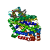
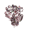
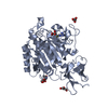
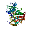
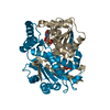
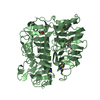
- PDB-6frv: Structure of the catalytic domain of Aspergillus niger Glucoamylase -
+ Open data

- Basic information
| Entry | Database: PDB / ID: 6frv | |||||||||
|---|---|---|---|---|---|---|---|---|---|---|
| Title | Structure of the catalytic domain of Aspergillus niger Glucoamylase | |||||||||
 Components Components | Glucoamylase | |||||||||
 Keywords Keywords | HYDROLASE / glycosylation / starch degradation / glycoside hydrolase | |||||||||
| Function / homology |  Function and homology information Function and homology informationglucan 1,4-alpha-glucosidase / polysaccharide metabolic process / glucan 1,4-alpha-glucosidase activity / starch binding / fungal-type vacuole / polysaccharide catabolic process / endoplasmic reticulum Similarity search - Function | |||||||||
| Biological species |  | |||||||||
| Method |  X-RAY DIFFRACTION / SYNCHROTRON / MOLECULAR REPLACEMENT / Resolution: 2.3 Å X-RAY DIFFRACTION / SYNCHROTRON / MOLECULAR REPLACEMENT / Resolution: 2.3 Å | |||||||||
 Authors Authors | Roth, C. / Moroz, O.V. / Ariza, A. / Friis, E.P. / Davies, G.J. / Wilson, K.S. | |||||||||
 Citation Citation | Journal: Acta Crystallogr D Struct Biol / Year: 2018 Title: Structural insight into industrially relevant glucoamylases: flexible positions of starch-binding domains. Authors: Roth, C. / Moroz, O.V. / Ariza, A. / Skov, L.K. / Ayabe, K. / Davies, G.J. / Wilson, K.S. | |||||||||
| History |
|
- Structure visualization
| Structure viewer | Molecule: MolmilJmol/JSmol |
|---|
- Downloads & links
-Download
| PDBx/mmCIF format | 6frv.cif.gz | 108.7 KB | Display | PDBx/mmCIF format |
|---|---|---|---|---|
| PDB format | pdb6frv.ent.gz | 78.7 KB | Display | PDB format |
| PDBx/mmJSON format | 6frv.json.gz | Tree view | PDBx/mmJSON format | |
| Others |  Other downloads Other downloads |
-Validation report
| Arichive directory | https://data.pdbj.org/pub/pdb/validation_reports/fr/6frvftp://data.pdbj.org/pub/pdb/validation_reports/fr/6frv | HTTPS FTP |
|---|
-Related structure data
| Related structure data | 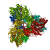 6fhvC 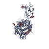 6fhwC 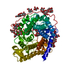 1agmS S: Starting model for refinement C: citing same article ( |
|---|---|
| Similar structure data |


-Links
 PDBj
PDBj


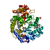
- Assembly
| Deposited unit | 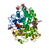
| ||||||||
|---|---|---|---|---|---|---|---|---|---|
| 1 |
| ||||||||
| Unit cell |
|
-Components
| #1: Protein | Mass: 65823.180 Da / Num. of mol.: 1 Source method: isolated from a genetically manipulated source Source: (gene. exp.) | ||||
|---|---|---|---|---|---|
| #2: Polysaccharide | 2-acetamido-2-deoxy-beta-D-glucopyranose-(1-4)-2-acetamido-2-deoxy-beta-D-glucopyranose Source method: isolated from a genetically manipulated source | ||||
| #3: Sugar | ChemComp-NAG /   Type: D-saccharide, beta linking / Mass: 221.208 Da / Num. of mol.: 1 Type: D-saccharide, beta linking / Mass: 221.208 Da / Num. of mol.: 1Source method: isolated from a genetically manipulated source Formula: C8H15NO6 | ||||
| #4: Sugar |   Type: D-saccharide, alpha linking / Mass: 180.156 Da / Num. of mol.: 2 / Source method: isolated from a natural source / Formula: C6H12O6 Type: D-saccharide, alpha linking / Mass: 180.156 Da / Num. of mol.: 2 / Source method: isolated from a natural source / Formula: C6H12O6#5: Water | ChemComp-HOH / |  Mass: 18.015 Da / Num. of mol.: 27 / Source method: isolated from a natural source / Formula: H2O Mass: 18.015 Da / Num. of mol.: 27 / Source method: isolated from a natural source / Formula: H2OHas protein modification | Y | |
-Experimental details
-Experiment
| Experiment | Method: X-RAY DIFFRACTION / Number of used crystals: 1 |
|---|
- Sample preparation
| Crystal | Density Matthews: 2.11 Å3/Da / Density % sol: 41.76 % |
|---|---|
| Crystal grow | Temperature: 292 K / Method: vapor diffusion, hanging drop / Details: PEG 3350, Hepes |
-Data collection
| Diffraction | Mean temperature: 100 K |
|---|---|
| Diffraction source | Source: SYNCHROTRON / Site: ESRF  / Beamline: ID14-2 / Wavelength: 0.933 Å / Beamline: ID14-2 / Wavelength: 0.933 Å |
| Detector | Type: ADSC QUANTUM 210 / Detector: CCD / Date: Dec 18, 2009 |
| Radiation | Protocol: SINGLE WAVELENGTH / Monochromatic (M) / Laue (L): M / Scattering type: x-ray |
| Radiation wavelength | Wavelength: 0.933 Å / Relative weight: 1 |
| Reflection | Resolution: 2.3→60 Å / Num. obs: 19454 / % possible obs: 99.5 % / Redundancy: 7.1 % / Rrim(I) all: 0.19 / Net I/σ(I): 6.3 |
| Reflection shell | Resolution: 2.3→2.38 Å / Num. unique obs: 1870 / Rrim(I) all: 1.516 |
- Processing
| Software |
| |||||||||||||||||||||||||||||||||||||||||||||||||||||||||||||||||||||||||||
|---|---|---|---|---|---|---|---|---|---|---|---|---|---|---|---|---|---|---|---|---|---|---|---|---|---|---|---|---|---|---|---|---|---|---|---|---|---|---|---|---|---|---|---|---|---|---|---|---|---|---|---|---|---|---|---|---|---|---|---|---|---|---|---|---|---|---|---|---|---|---|---|---|---|---|---|---|
| Refinement | Method to determine structure: MOLECULAR REPLACEMENT Starting model: 1agm Resolution: 2.3→59.67 Å / Cor.coef. Fo:Fc: 0.937 / Cor.coef. Fo:Fc free: 0.862 / Cross valid method: THROUGHOUT / σ(F): 0 / ESU R: 0.567 / ESU R Free: 0.357 Details: HYDROGENS HAVE BEEN ADDED IN THE RIDING POSITIONS U VALUES : REFINED INDIVIDUALLY
| |||||||||||||||||||||||||||||||||||||||||||||||||||||||||||||||||||||||||||
| Solvent computation | Ion probe radii: 0.8 Å / Shrinkage radii: 0.8 Å / VDW probe radii: 1.2 Å | |||||||||||||||||||||||||||||||||||||||||||||||||||||||||||||||||||||||||||
| Displacement parameters | Biso max: 125.17 Å2 / Biso mean: 56.096 Å2 / Biso min: 25.05 Å2
| |||||||||||||||||||||||||||||||||||||||||||||||||||||||||||||||||||||||||||
| Refinement step | Cycle: final / Resolution: 2.3→59.67 Å
| |||||||||||||||||||||||||||||||||||||||||||||||||||||||||||||||||||||||||||
| Refine LS restraints |
| |||||||||||||||||||||||||||||||||||||||||||||||||||||||||||||||||||||||||||
| LS refinement shell | Resolution: 2.3→2.36 Å / Total num. of bins used: 20
|
