 Movie
Movie Controller

[English] 日本語
 Yorodumi
- PDB-6pwq: Crystal structure of Levansucrase from Bacillus subtilis mutant S... -
+ Open data

- Basic information
| Entry | Database: PDB / ID: 6pwq | ||||||||||||
|---|---|---|---|---|---|---|---|---|---|---|---|---|---|
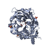
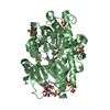
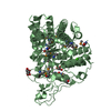
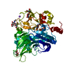
| Title | Crystal structure of Levansucrase from Bacillus subtilis mutant S164A at 2.6 A | ||||||||||||
 Components Components | Glycoside hydrolase family 68 protein | ||||||||||||
 Keywords Keywords | TRANSFERASE / Levansucrase / Glycoside hydrolase / Levan / Fructose polymers | ||||||||||||
| Function / homology |  Function and homology information Function and homology informationlevansucrase / levansucrase activity / carbohydrate utilization / extracellular region / metal ion binding Similarity search - Function | ||||||||||||
| Biological species |  | ||||||||||||
| Method |  X-RAY DIFFRACTION / SYNCHROTRON / MOLECULAR REPLACEMENT / Resolution: 2.6 Å X-RAY DIFFRACTION / SYNCHROTRON / MOLECULAR REPLACEMENT / Resolution: 2.6 Å | ||||||||||||
 Authors Authors | Diaz-Vilchis, A. / Rodriguez-Alegria, M.E. / Ortiz-Soto, M.E. / Rudino-Pinera, E. / Lopez-Munguia, A. | ||||||||||||
| Funding support |  Mexico, Mexico,  Germany, 3items Germany, 3items
| ||||||||||||
 Citation Citation | Journal: Int.J.Biol.Macromol. / Year: 2020 Title: Implications of the mutation S164A on Bacillus subtilis levansucrase product specificity and insights into protein interactions acting upon levan synthesis. Authors: Ortiz-Soto, M.E. / Porras-Dominguez, J.R. / Rodriguez-Alegria, M.E. / Morales-Moreno, L.A. / Diaz-Vilchis, A. / Rudino-Pinera, E. / Beltran-Hernandez, N.E. / Rivera, H.M. / Seibel, J. / Lopez Munguia, A. | ||||||||||||
| History |
|
- Structure visualization
| Structure viewer | Molecule: MolmilJmol/JSmol |
|---|
- Downloads & links
-Download
| PDBx/mmCIF format | 6pwq.cif.gz | 195.1 KB | Display | PDBx/mmCIF format |
|---|---|---|---|---|
| PDB format | pdb6pwq.ent.gz | 153.5 KB | Display | PDB format |
| PDBx/mmJSON format | 6pwq.json.gz | Tree view | PDBx/mmJSON format | |
| Others |  Other downloads Other downloads |
-Validation report
| Arichive directory | https://data.pdbj.org/pub/pdb/validation_reports/pw/6pwqftp://data.pdbj.org/pub/pdb/validation_reports/pw/6pwq | HTTPS FTP |
|---|
-Related structure data
| Related structure data | 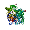 1oygS S: Starting model for refinement |
|---|---|
| Similar structure data |


-Links
 PDBj
PDBj- Assembly
| Deposited unit | 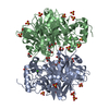
| ||||||||
|---|---|---|---|---|---|---|---|---|---|
| 1 | 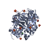
| ||||||||
| 2 | 
| ||||||||
| Unit cell |
|
-Components
-Protein , 1 types, 2 molecules AB
| #1: Protein | Mass: 52446.844 Da / Num. of mol.: 2 / Mutation: S164A Source method: isolated from a genetically manipulated source Details: Calcium Sulfate Tetraethylene Glycol Glycerol / Source: (gene. exp.) |
|---|
-Non-polymers , 5 types, 216 molecules 


| #2: Chemical |  Mass: 40.078 Da / Num. of mol.: 2 / Source method: obtained synthetically / Formula: Ca / Feature type: SUBJECT OF INVESTIGATION Mass: 40.078 Da / Num. of mol.: 2 / Source method: obtained synthetically / Formula: Ca / Feature type: SUBJECT OF INVESTIGATION#3: Chemical | ChemComp-SO4 /  Mass: 96.063 Da / Num. of mol.: 20 / Source method: obtained synthetically / Formula: SO4 / Feature type: SUBJECT OF INVESTIGATION Mass: 96.063 Da / Num. of mol.: 20 / Source method: obtained synthetically / Formula: SO4 / Feature type: SUBJECT OF INVESTIGATION#4: Chemical |  Mass: 194.226 Da / Num. of mol.: 2 / Source method: obtained synthetically / Formula: C8H18O5 / Feature type: SUBJECT OF INVESTIGATION / Comment: precipitant*YM Mass: 194.226 Da / Num. of mol.: 2 / Source method: obtained synthetically / Formula: C8H18O5 / Feature type: SUBJECT OF INVESTIGATION / Comment: precipitant*YM#5: Chemical | ChemComp-GOL /  Mass: 92.094 Da / Num. of mol.: 5 / Source method: obtained synthetically / Formula: C3H8O3 / Feature type: SUBJECT OF INVESTIGATION Mass: 92.094 Da / Num. of mol.: 5 / Source method: obtained synthetically / Formula: C3H8O3 / Feature type: SUBJECT OF INVESTIGATION#6: Water | ChemComp-HOH / | Mass: 18.015 Da / Num. of mol.: 187 / Source method: isolated from a natural source / Formula: H2O |
|---|
-Details
| Has ligand of interest | Y |
|---|
-Experimental details
-Experiment
| Experiment | Method: X-RAY DIFFRACTION / Number of used crystals: 1 |
|---|
- Sample preparation
| Crystal | Density Matthews: 3.1 Å3/Da / Density % sol: 60.36 % / Description: irregular octahedron |
|---|---|
| Crystal grow | Temperature: 291 K / Method: vapor diffusion, sitting drop / pH: 5 Details: 3.2 M ammonium sulfate and 100 mM citric acid/sodium hydroxide, pH 5.0. |
-Data collection
| Diffraction | Mean temperature: 100 K / Serial crystal experiment: N |
|---|---|
| Diffraction source | Source: SYNCHROTRON / Site: APS  / Beamline: 19-BM / Wavelength: 1.02 Å / Beamline: 19-BM / Wavelength: 1.02 Å |
| Detector | Type: ADSC QUANTUM 210r / Detector: CCD / Date: Oct 17, 2018 |
| Radiation | Protocol: SINGLE WAVELENGTH / Monochromatic (M) / Laue (L): M / Scattering type: x-ray |
| Radiation wavelength | Wavelength: 1.02 Å / Relative weight: 1 |
| Reflection | Resolution: 2.6→25 Å / Num. obs: 38955 / % possible obs: 99.9 % / Observed criterion σ(I): 3.1 / Redundancy: 6.8 % / Biso Wilson estimate: 45.9 Å2 / CC1/2: 0.99 / Rmerge(I) obs: 0.074 / Net I/σ(I): 14.5 |
| Reflection shell | Resolution: 2.6→2.71 Å / Redundancy: 6.1 % / Rmerge(I) obs: 0.51 / Mean I/σ(I) obs: 3.1 / Num. unique obs: 3808 / CC1/2: 0.81 / % possible all: 100 |
- Processing
| Software |
| |||||||||||||||||||||||||||||||||||||||||||||||||||||||||||||||||||||||||||
|---|---|---|---|---|---|---|---|---|---|---|---|---|---|---|---|---|---|---|---|---|---|---|---|---|---|---|---|---|---|---|---|---|---|---|---|---|---|---|---|---|---|---|---|---|---|---|---|---|---|---|---|---|---|---|---|---|---|---|---|---|---|---|---|---|---|---|---|---|---|---|---|---|---|---|---|---|
| Refinement | Method to determine structure: MOLECULAR REPLACEMENT Starting model: 1OYG Resolution: 2.6→19.9782 Å / SU ML: 0.27 / Cross valid method: FREE R-VALUE / σ(F): 1.36 / Phase error: 23.43
| |||||||||||||||||||||||||||||||||||||||||||||||||||||||||||||||||||||||||||
| Solvent computation | Shrinkage radii: 0.9 Å / VDW probe radii: 1.11 Å | |||||||||||||||||||||||||||||||||||||||||||||||||||||||||||||||||||||||||||
| Displacement parameters | Biso max: 108.68 Å2 / Biso mean: 48.7225 Å2 / Biso min: 29.13 Å2 | |||||||||||||||||||||||||||||||||||||||||||||||||||||||||||||||||||||||||||
| Refinement step | Cycle: final / Resolution: 2.6→19.9782 Å
| |||||||||||||||||||||||||||||||||||||||||||||||||||||||||||||||||||||||||||
| Refine LS restraints |
| |||||||||||||||||||||||||||||||||||||||||||||||||||||||||||||||||||||||||||
| LS refinement shell | Refine-ID: X-RAY DIFFRACTION / Rfactor Rfree error: 0 / % reflection obs: 100 %
|
