- PDB-4u90: GephE in complex with PEG crosslinked GABA receptor alpha3 subuni... -
+
Open data
ID or keywords:
Loading...
-
Basic information
Entry
Database: PDB / ID: 4u90
Title
GephE in complex with PEG crosslinked GABA receptor alpha3 subunit derived dimeric peptide
Components
Gamma-aminobutyric acid receptor subunit alpha-3
Gephyrin
Keywords
TRANSFER PROTEIN / STRUCTURAL PROTEIN / Inhibitory synapse / Scaffolding protein / GABA type A receptor / transferase / transfer protein - structural protein complex
Function / homology
Function and homology information
GABA receptor activation / Molybdenum cofactor biosynthesis / glycine receptor clustering / molybdopterin cofactor biosynthetic process / establishment of synaptic specificity at neuromuscular junction / molybdopterin adenylyltransferase activity / nitrate reductase activity / molybdopterin molybdotransferase activity / molybdopterin adenylyltransferase / molybdopterin molybdotransferase ...GABA receptor activation / Molybdenum cofactor biosynthesis / glycine receptor clustering / molybdopterin cofactor biosynthetic process / establishment of synaptic specificity at neuromuscular junction / molybdopterin adenylyltransferase activity / nitrate reductase activity / molybdopterin molybdotransferase activity / molybdopterin adenylyltransferase / molybdopterin molybdotransferase / gamma-aminobutyric acid receptor clustering / postsynaptic specialization / inhibitory synapse / Mo-molybdopterin cofactor biosynthetic process / auditory behavior / glycinergic synapse / molybdopterin cofactor binding / GABA-gated chloride ion channel activity / GABA-A receptor complex / inhibitory synapse assembly / GABA-A receptor activity / postsynaptic specialization, intracellular component / response to metal ion / postsynaptic specialization membrane / gamma-aminobutyric acid signaling pathway / neurotransmitter receptor localization to postsynaptic specialization membrane / synaptic transmission, GABAergic / chloride channel complex / protein targeting / presynaptic active zone membrane / synapse assembly / dendrite membrane / ligand-gated monoatomic ion channel activity involved in regulation of presynaptic membrane potential / chloride transmembrane transport / synaptic membrane / establishment of protein localization / transmitter-gated monoatomic ion channel activity involved in regulation of postsynaptic membrane potential / tubulin binding / response to lead ion / GABA-ergic synapse / cytoplasmic side of plasma membrane / molecular adaptor activity / dendritic spine / cytoskeleton / chemical synaptic transmission / protein-macromolecule adaptor activity / postsynaptic membrane / postsynaptic density / postsynapse / signaling receptor binding / neuronal cell body / synapse / dendrite / ATP binding / metal ion binding / identical protein binding / cytoplasm / cytosol Similarity search - Function
Resolution: 2→20 Å / Cor.coef. Fo:Fc: 0.969 / Cor.coef. Fo:Fc free: 0.951 / SU B: 8.411 / SU ML: 0.115 / Cross valid method: THROUGHOUT / ESU R: 0.151 / ESU R Free: 0.143 / Stereochemistry target values: MAXIMUM LIKELIHOOD / Details: HYDROGENS HAVE BEEN ADDED IN THE RIDING POSITIONS
Rfactor
Num. reflection
% reflection
Selection details
Rfree
0.21103
1694
4.9 %
RANDOM
Rwork
0.16655
-
-
-
obs
0.16878
32616
99.67 %
-
Solvent computation
Ion probe radii: 0.8 Å / Shrinkage radii: 0.8 Å / VDW probe radii: 1.2 Å / Solvent model: BABINET MODEL WITH MASK
 Movie
Movie Controller

 Yorodumi
 Open data
 Basic information
 Components
 Keywords
 Function and homology information
 X-RAY DIFFRACTION /
  Authors
 Germany, 1items
  Citation
 Structure visualization
 Downloads & links
 Other downloads


 PDBj
PDBj


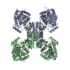
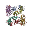
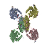
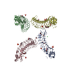
 Assembly
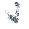


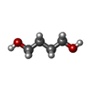
 Mass: 90.121 Da / Num. of mol.: 1 / Source method: obtained synthetically / Formula: C4H10O2
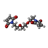
 Mass: 308.287 Da / Num. of mol.: 1 / Source method: obtained synthetically / Formula: C14H16N2O6
 Mass: 18.015 Da / Num. of mol.: 255 / Source method: isolated from a natural source / Formula: H2O
 Sample preparation
 / Beamline: ID23-1 / Wavelength: 0.91 Å
 Processing