 Movie
Movie Controller

[English] 日本語
 Yorodumi
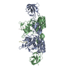
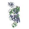
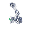
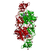
- PDB-4pd1: Structure of gephyrin E domain with Glycine-beta receptor peptide -
+ Open data

- Basic information
| Entry | Database: PDB / ID: 4pd1 | ||||||
|---|---|---|---|---|---|---|---|
| Title | Structure of gephyrin E domain with Glycine-beta receptor peptide | ||||||
 Components Components |
| ||||||
 Keywords Keywords | STRUCTURAL PROTEIN/TRANSPORT PROTEIN / Scaffolding protein / Neurotransmitter receptor anchoring protein / Molybdenum cofactor biosynthesis / STRUCTURAL PROTEIN-SIGNALING PROTEIN complex / STRUCTURAL PROTEIN-TRANSPORT PROTEIN complex | ||||||
| Function / homology |  Function and homology information Function and homology informationNeurotransmitter receptors and postsynaptic signal transmission / Molybdenum cofactor biosynthesis / glycine receptor clustering / molybdopterin cofactor biosynthetic process / acrosome reaction / establishment of synaptic specificity at neuromuscular junction / glycine-gated chloride channel complex / synaptic transmission, glycinergic / molybdopterin adenylyltransferase activity / molybdopterin adenylyltransferase ...Neurotransmitter receptors and postsynaptic signal transmission / Molybdenum cofactor biosynthesis / glycine receptor clustering / molybdopterin cofactor biosynthetic process / acrosome reaction / establishment of synaptic specificity at neuromuscular junction / glycine-gated chloride channel complex / synaptic transmission, glycinergic / molybdopterin adenylyltransferase activity / molybdopterin adenylyltransferase / nitrate reductase activity / molybdopterin molybdotransferase activity / molybdopterin molybdotransferase / gamma-aminobutyric acid receptor clustering / postsynaptic specialization / extracellularly glycine-gated ion channel activity / righting reflex / extracellularly glycine-gated chloride channel activity / inhibitory synapse / Mo-molybdopterin cofactor biosynthetic process / glycinergic synapse / molybdopterin cofactor binding / postsynaptic specialization, intracellular component / excitatory extracellular ligand-gated monoatomic ion channel activity / adult walking behavior / response to metal ion / neuromuscular process / neurotransmitter receptor localization to postsynaptic specialization membrane / glycine binding / synaptic transmission, GABAergic / startle response / protein targeting / monoatomic ion transport / visual perception / synapse assembly / chloride transmembrane transport / synaptic membrane / establishment of protein localization / neuropeptide signaling pathway / transmitter-gated monoatomic ion channel activity involved in regulation of postsynaptic membrane potential / regulation of membrane potential / tubulin binding / GABA-ergic synapse / cytoplasmic side of plasma membrane / transmembrane signaling receptor activity / nervous system development / molecular adaptor activity / dendritic spine / cytoskeleton / chemical synaptic transmission / perikaryon / protein-macromolecule adaptor activity / postsynaptic membrane / postsynaptic density / postsynapse / signaling receptor binding / neuronal cell body / dendrite / protein-containing complex binding / ATP binding / membrane / metal ion binding / identical protein binding / plasma membrane / cytoplasm / cytosol Similarity search - Function | ||||||
| Biological species |  | ||||||
| Method |  X-RAY DIFFRACTION / SYNCHROTRON / MOLECULAR REPLACEMENT / Resolution: 1.975 Å X-RAY DIFFRACTION / SYNCHROTRON / MOLECULAR REPLACEMENT / Resolution: 1.975 Å | ||||||
 Authors Authors | Kasaragod, V.B. / Maric, H.M. / Schindelin, H. | ||||||
| Funding support |  Germany, 1items Germany, 1items
| ||||||
 Citation Citation | Journal: Acs Chem.Biol. / Year: 2014 Title: Modulation of gephyrin-glycine receptor affinity by multivalency. Authors: Maric, H.M. / Kasaragod, V.B. / Schindelin, H. | ||||||
| History |
|
- Structure visualization
| Structure viewer | Molecule: MolmilJmol/JSmol |
|---|
- Downloads & links
-Download
| PDBx/mmCIF format | 4pd1.cif.gz | 250.2 KB | Display | PDBx/mmCIF format |
|---|---|---|---|---|
| PDB format | pdb4pd1.ent.gz | 205.5 KB | Display | PDB format |
| PDBx/mmJSON format | 4pd1.json.gz | Tree view | PDBx/mmJSON format | |
| Others |  Other downloads Other downloads |
-Validation report
| Arichive directory | https://data.pdbj.org/pub/pdb/validation_reports/pd/4pd1ftp://data.pdbj.org/pub/pdb/validation_reports/pd/4pd1 | HTTPS FTP |
|---|
-Related structure data
| Related structure data |  4pd0C  2fu3S C: citing same article ( S: Starting model for refinement |
|---|---|
| Similar structure data |


-Links
 PDBj
PDBj

- Assembly
| Deposited unit | 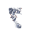
| |||||||||||||||
|---|---|---|---|---|---|---|---|---|---|---|---|---|---|---|---|---|
| 1 | 
| |||||||||||||||
| Unit cell |
| |||||||||||||||
| Components on special symmetry positions |
|
-Components
| #1: Protein | Mass: 45652.395 Da / Num. of mol.: 1 / Fragment: E-domain (UNP residues 350-768) Source method: isolated from a genetically manipulated source Source: (gene. exp.)  References: UniProt: Q03555, molybdopterin adenylyltransferase, molybdopterin molybdotransferase | ||||
|---|---|---|---|---|---|
| #2: Protein/peptide | Mass: 1682.850 Da / Num. of mol.: 1 / Fragment: UNP residues 419-433 / Source method: obtained synthetically / Source: (synth.) | ||||
| #3: Chemical | ChemComp-GOL /   Mass: 92.094 Da / Num. of mol.: 8 / Source method: obtained synthetically / Formula: C3H8O3 Mass: 92.094 Da / Num. of mol.: 8 / Source method: obtained synthetically / Formula: C3H8O3#4: Chemical | ChemComp-ACT / |   Mass: 59.044 Da / Num. of mol.: 1 / Source method: obtained synthetically / Formula: C2H3O2 Mass: 59.044 Da / Num. of mol.: 1 / Source method: obtained synthetically / Formula: C2H3O2#5: Water | ChemComp-HOH / |  Mass: 18.015 Da / Num. of mol.: 174 / Source method: isolated from a natural source / Formula: H2O Mass: 18.015 Da / Num. of mol.: 174 / Source method: isolated from a natural source / Formula: H2O |
-Experimental details
-Experiment
| Experiment | Method: X-RAY DIFFRACTION / Number of used crystals: 1 |
|---|
- Sample preparation
| Crystal | Density Matthews: 2.66 Å3/Da / Density % sol: 53.75 % |
|---|---|
| Crystal grow | Temperature: 293 K / Method: vapor diffusion, sitting drop / pH: 4.5 Details: 0.1 M sodium citrate, pH 4.5, 28-34 % 2-methyl-2-4-pentanediol |
-Data collection
| Diffraction | Mean temperature: 100 K |
|---|---|
| Diffraction source | Source: SYNCHROTRON / Site: BESSY / Beamline: 14.1 / Wavelength: 0.9184 Å |
| Detector | Type: MARMOSAIC 225 mm CCD / Detector: CCD / Date: Jul 29, 2011 |
| Radiation | Protocol: SINGLE WAVELENGTH / Monochromatic (M) / Laue (L): M / Scattering type: x-ray |
| Radiation wavelength | Wavelength: 0.9184 Å / Relative weight: 1 |
| Reflection | Resolution: 1.975→36.472 Å / Num. obs: 35783 / % possible obs: 100 % / Redundancy: 14.7 % / Rmerge(I) obs: 0.111 / Net I/σ(I): 17.7 |
- Processing
| Software | Name: PHENIX / Version: (phenix.refine: 1.8_1069) / Classification: refinement | ||||||||||||||||||||||||||||||||||||||||||||||||||||||||||||||||||||||||||||||||||||||||||||||||||||||||||||||||||||||||||||||||||||||||||||||||||||||||||||||||||||||||||||||||||||||||||||||||||||||||||||||||||||||||||||||||||||||||||||||||||||||||||||||||||||||||||||||||||||||||||||||||||||||||||||
|---|---|---|---|---|---|---|---|---|---|---|---|---|---|---|---|---|---|---|---|---|---|---|---|---|---|---|---|---|---|---|---|---|---|---|---|---|---|---|---|---|---|---|---|---|---|---|---|---|---|---|---|---|---|---|---|---|---|---|---|---|---|---|---|---|---|---|---|---|---|---|---|---|---|---|---|---|---|---|---|---|---|---|---|---|---|---|---|---|---|---|---|---|---|---|---|---|---|---|---|---|---|---|---|---|---|---|---|---|---|---|---|---|---|---|---|---|---|---|---|---|---|---|---|---|---|---|---|---|---|---|---|---|---|---|---|---|---|---|---|---|---|---|---|---|---|---|---|---|---|---|---|---|---|---|---|---|---|---|---|---|---|---|---|---|---|---|---|---|---|---|---|---|---|---|---|---|---|---|---|---|---|---|---|---|---|---|---|---|---|---|---|---|---|---|---|---|---|---|---|---|---|---|---|---|---|---|---|---|---|---|---|---|---|---|---|---|---|---|---|---|---|---|---|---|---|---|---|---|---|---|---|---|---|---|---|---|---|---|---|---|---|---|---|---|---|---|---|---|---|---|---|---|---|---|---|---|---|---|---|---|---|---|---|---|---|---|---|---|---|---|---|---|---|---|---|---|---|---|---|---|---|---|---|---|---|---|---|---|---|---|---|---|---|---|---|---|---|---|---|---|---|
| Refinement | Method to determine structure: MOLECULAR REPLACEMENT Starting model: 2FU3 Resolution: 1.975→36.472 Å / SU ML: 0.17 / Cross valid method: FREE R-VALUE / σ(F): 1.36 / Phase error: 17.82 / Stereochemistry target values: ML
| ||||||||||||||||||||||||||||||||||||||||||||||||||||||||||||||||||||||||||||||||||||||||||||||||||||||||||||||||||||||||||||||||||||||||||||||||||||||||||||||||||||||||||||||||||||||||||||||||||||||||||||||||||||||||||||||||||||||||||||||||||||||||||||||||||||||||||||||||||||||||||||||||||||||||||||
| Solvent computation | Shrinkage radii: 0.9 Å / VDW probe radii: 1.11 Å / Solvent model: FLAT BULK SOLVENT MODEL | ||||||||||||||||||||||||||||||||||||||||||||||||||||||||||||||||||||||||||||||||||||||||||||||||||||||||||||||||||||||||||||||||||||||||||||||||||||||||||||||||||||||||||||||||||||||||||||||||||||||||||||||||||||||||||||||||||||||||||||||||||||||||||||||||||||||||||||||||||||||||||||||||||||||||||||
| Refinement step | Cycle: LAST / Resolution: 1.975→36.472 Å
| ||||||||||||||||||||||||||||||||||||||||||||||||||||||||||||||||||||||||||||||||||||||||||||||||||||||||||||||||||||||||||||||||||||||||||||||||||||||||||||||||||||||||||||||||||||||||||||||||||||||||||||||||||||||||||||||||||||||||||||||||||||||||||||||||||||||||||||||||||||||||||||||||||||||||||||
| Refine LS restraints |
| ||||||||||||||||||||||||||||||||||||||||||||||||||||||||||||||||||||||||||||||||||||||||||||||||||||||||||||||||||||||||||||||||||||||||||||||||||||||||||||||||||||||||||||||||||||||||||||||||||||||||||||||||||||||||||||||||||||||||||||||||||||||||||||||||||||||||||||||||||||||||||||||||||||||||||||
| LS refinement shell |
| ||||||||||||||||||||||||||||||||||||||||||||||||||||||||||||||||||||||||||||||||||||||||||||||||||||||||||||||||||||||||||||||||||||||||||||||||||||||||||||||||||||||||||||||||||||||||||||||||||||||||||||||||||||||||||||||||||||||||||||||||||||||||||||||||||||||||||||||||||||||||||||||||||||||||||||
| Refinement TLS params. | Method: refined / Refine-ID: X-RAY DIFFRACTION
| ||||||||||||||||||||||||||||||||||||||||||||||||||||||||||||||||||||||||||||||||||||||||||||||||||||||||||||||||||||||||||||||||||||||||||||||||||||||||||||||||||||||||||||||||||||||||||||||||||||||||||||||||||||||||||||||||||||||||||||||||||||||||||||||||||||||||||||||||||||||||||||||||||||||||||||
| Refinement TLS group |
|
