| Entry | Database: PDB / ID: 1t3e
|
|---|
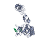
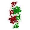
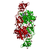
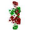
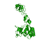
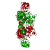
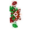
| Title | Structural basis of dynamic glycine receptor clustering |
|---|
 Components Components | - 49-mer fragment of Glycine receptor beta chain
- Gephyrin
|
|---|
 Keywords Keywords | STRUCTURAL PROTEIN/SIGNALING PROTEIN / alfa-beta / STRUCTURAL PROTEIN-SIGNALING PROTEIN COMPLEX |
|---|
| Function / homology |  Function and homology information Function and homology information
Neurotransmitter receptors and postsynaptic signal transmission / Molybdenum cofactor biosynthesis / glycine receptor clustering / molybdopterin cofactor biosynthetic process / acrosome reaction / establishment of synaptic specificity at neuromuscular junction / glycine-gated chloride channel complex / synaptic transmission, glycinergic / molybdopterin adenylyltransferase activity / molybdopterin adenylyltransferase ...Neurotransmitter receptors and postsynaptic signal transmission / Molybdenum cofactor biosynthesis / glycine receptor clustering / molybdopterin cofactor biosynthetic process / acrosome reaction / establishment of synaptic specificity at neuromuscular junction / glycine-gated chloride channel complex / synaptic transmission, glycinergic / molybdopterin adenylyltransferase activity / molybdopterin adenylyltransferase / nitrate reductase activity / molybdopterin molybdotransferase activity / molybdopterin molybdotransferase / gamma-aminobutyric acid receptor clustering / postsynaptic specialization / extracellularly glycine-gated ion channel activity / righting reflex / extracellularly glycine-gated chloride channel activity / inhibitory synapse / Mo-molybdopterin cofactor biosynthetic process / glycinergic synapse / molybdopterin cofactor binding / postsynaptic specialization, intracellular component / excitatory extracellular ligand-gated monoatomic ion channel activity / adult walking behavior / response to metal ion / neuromuscular process / neurotransmitter receptor localization to postsynaptic specialization membrane / glycine binding / synaptic transmission, GABAergic / startle response / protein targeting / monoatomic ion transport / visual perception / synapse assembly / chloride transmembrane transport / synaptic membrane / establishment of protein localization / neuropeptide signaling pathway / transmitter-gated monoatomic ion channel activity involved in regulation of postsynaptic membrane potential / regulation of membrane potential / tubulin binding / GABA-ergic synapse / cytoplasmic side of plasma membrane / transmembrane signaling receptor activity / nervous system development / molecular adaptor activity / dendritic spine / cytoskeleton / chemical synaptic transmission / perikaryon / protein-macromolecule adaptor activity / postsynaptic membrane / postsynaptic density / postsynapse / signaling receptor binding / neuronal cell body / dendrite / protein-containing complex binding / ATP binding / membrane / metal ion binding / identical protein binding / plasma membrane / cytoplasm / cytosolSimilarity search - Function : / Glycine receptor beta / : / Molybdopterin biosynthesis moeA protein; domain 3 / Molybdopterin biosynthesis moea protein, domain 3. / Beta-clip / MoeA, C-terminal, domain IV / Molybdopterin biosynthesis moea protein, domain 2 / Molybdenum cofactor biosynthesis proteins signature 2. / Molybdenum cofactor biosynthesis proteins signature 1. ...: / Glycine receptor beta / : / Molybdopterin biosynthesis moeA protein; domain 3 / Molybdopterin biosynthesis moea protein, domain 3. / Beta-clip / MoeA, C-terminal, domain IV / Molybdopterin biosynthesis moea protein, domain 2 / Molybdenum cofactor biosynthesis proteins signature 2. / Molybdenum cofactor biosynthesis proteins signature 1. / MoeA, N-terminal and linker domain / MoeA, C-terminal, domain IV / MoeA, N-terminal and linker domain superfamily / MoeA, C-terminal, domain IV superfamily / Molybdopterin biosynthesis protein MoeA-like / MoeA N-terminal region (domain I and II) / MoeA C-terminal region (domain IV) / Molybdopterin biosynthesis moea protein, domain 2 / Molybdenum cofactor biosynthesis, conserved site / MoaB/Mog-like domain / Molybdenum Cofactor Biosythetic Enzyme; Chain A / MoaB/Mog domain / MoaB/Mog-like domain superfamily / Probable molybdopterin binding domain / Probable molybdopterin binding domain / Neurotransmitter-gated ion-channel transmembrane domain / Neurotransmitter-gated ion-channel, conserved site / Neurotransmitter-gated ion-channel transmembrane region / Neurotransmitter-gated ion-channels signature. / Neurotransmitter-gated ion-channel transmembrane domain superfamily / Neuronal acetylcholine receptor / Neurotransmitter-gated ion-channel / Neurotransmitter-gated ion-channel ligand-binding domain / Neurotransmitter-gated ion-channel ligand-binding domain superfamily / Neurotransmitter-gated ion-channel ligand binding domain / Beta Complex / Alpha-Beta Complex / Beta Barrel / 3-Layer(aba) Sandwich / Mainly Beta / Alpha BetaSimilarity search - Domain/homology |
|---|
| Biological species |   Rattus norvegicus (Norway rat) Rattus norvegicus (Norway rat) |
|---|
| Method |  X-RAY DIFFRACTION / SYNCHROTRON / MOLECULAR REPLACEMENT / Resolution: 3.25 Å X-RAY DIFFRACTION / SYNCHROTRON / MOLECULAR REPLACEMENT / Resolution: 3.25 Å |
|---|
 Authors Authors | Sola, M. / Bavro, V.N. / Timmins, J. / Franz, T. / Ricard-Blum, S. / Schoehn, G. / Ruigrok, R.W.H. / Paarmann, I. / Saiyed, T. / O'Sullivan, G.A. |
|---|
 Citation Citation | Journal: Embo J. / Year: 2004
Title: Structural basis of dynamic glycine receptor clustering by gephyrin
Authors: Sola, M. / Bavro, V.N. / Timmins, J. / Franz, T. / Ricard-Blum, S. / Schoehn, G. / Ruigrok, R.W.H. / Paarmann, I. / Saiyed, T. / O'Sullivan, G.A. / Schmitt, B. / Betz, H. / Weissenhorn, W. |
|---|
| History | | Deposition | Apr 26, 2004 | Deposition site: RCSB / Processing site: RCSB |
|---|
| Revision 1.0 | Jul 27, 2004 | Provider: repository / Type: Initial release |
|---|
| Revision 1.1 | Apr 30, 2008 | Group: Version format compliance |
|---|
| Revision 1.2 | Jul 13, 2011 | Group: Advisory / Derived calculations ...Advisory / Derived calculations / Refinement description / Version format compliance |
|---|
| Revision 1.3 | Feb 14, 2024 | Group: Data collection / Database references ...Data collection / Database references / Derived calculations / Refinement description
Category: chem_comp_atom / chem_comp_bond ...chem_comp_atom / chem_comp_bond / database_2 / struct_ncs_dom_lim / struct_site
Item: _database_2.pdbx_DOI / _database_2.pdbx_database_accession ..._database_2.pdbx_DOI / _database_2.pdbx_database_accession / _struct_ncs_dom_lim.beg_auth_comp_id / _struct_ncs_dom_lim.end_auth_comp_id / _struct_site.pdbx_auth_asym_id / _struct_site.pdbx_auth_comp_id / _struct_site.pdbx_auth_seq_id |
|---|
| Revision 1.4 | Apr 3, 2024 | Group: Refinement description / Category: pdbx_initial_refinement_model |
|---|
|
|---|
 Movie
Movie Controller


 Open data
 Basic information
 Structure visualization
 Downloads & links
 Other downloads
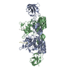


 PDBj
PDBj

 Assembly