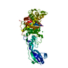
 Movie
Movie Controller


+ Open data

- Basic information
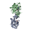
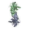
| Entry | Database: PDB / ID: 4u16 | ||||||
|---|---|---|---|---|---|---|---|
| Title | M3-mT4L receptor bound to NMS | ||||||
 Components Components | Muscarinic acetylcholine receptor M3,Lysozyme,Muscarinic acetylcholine receptor M3 | ||||||
 Keywords Keywords | MEMBRANE PROTEIN/INHIBITOR / GPCR Crystallography T4 lysozyme / MEMBRANE PROTEIN-INHIBITOR complex | ||||||
| Function / homology |  Function and homology information Function and homology informationnegative regulation of heart rate by acetylcholine / G protein-coupled acetylcholine receptor binding / Muscarinic acetylcholine receptors / quaternary ammonium group binding / saliva secretion / phospholipase C-activating G protein-coupled acetylcholine receptor signaling pathway / G protein-coupled acetylcholine receptor activity / response to acetylcholine / positive regulation of vascular associated smooth muscle contraction / regulation of smooth muscle contraction ...negative regulation of heart rate by acetylcholine / G protein-coupled acetylcholine receptor binding / Muscarinic acetylcholine receptors / quaternary ammonium group binding / saliva secretion / phospholipase C-activating G protein-coupled acetylcholine receptor signaling pathway / G protein-coupled acetylcholine receptor activity / response to acetylcholine / positive regulation of vascular associated smooth muscle contraction / regulation of smooth muscle contraction / positive regulation of smooth muscle contraction / adenylate cyclase-inhibiting G protein-coupled acetylcholine receptor signaling pathway / acetylcholine binding / G alpha (q) signalling events / acetylcholine receptor signaling pathway / synaptic transmission, cholinergic / ligand-gated ion channel signaling pathway / smooth muscle contraction / positive regulation of vasoconstriction / asymmetric synapse / G protein-coupled receptor signaling pathway, coupled to cyclic nucleotide second messenger / viral release from host cell by cytolysis / peptidoglycan catabolic process / basal plasma membrane / axon terminus / calcium-mediated signaling / postsynaptic density membrane / cell wall macromolecule catabolic process / positive regulation of insulin secretion / lysozyme / lysozyme activity / G protein-coupled acetylcholine receptor signaling pathway / presynaptic membrane / basolateral plasma membrane / host cell cytoplasm / chemical synaptic transmission / defense response to bacterium / synapse / dendrite / endoplasmic reticulum membrane / glutamatergic synapse / plasma membrane Similarity search - Function | ||||||
| Biological species |   Enterobacteria phage T4 (virus) Enterobacteria phage T4 (virus) | ||||||
| Method |  X-RAY DIFFRACTION / SYNCHROTRON / MOLECULAR REPLACEMENT / Resolution: 3.7 Å X-RAY DIFFRACTION / SYNCHROTRON / MOLECULAR REPLACEMENT / Resolution: 3.7 Å | ||||||
 Authors Authors | Thorsen, T.S. / Matt, R. / Weis, W.I. / Kobilka, B. | ||||||
 Citation Citation | Journal: Structure / Year: 2014 Title: Modified T4 Lysozyme Fusion Proteins Facilitate G Protein-Coupled Receptor Crystallogenesis. Authors: Thorsen, T.S. / Matt, R. / Weis, W.I. / Kobilka, B.K. | ||||||
| History |
|
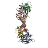
- Structure visualization
| Structure viewer | Molecule: MolmilJmol/JSmol |
|---|
- Downloads & links
-Download
| PDBx/mmCIF format | 4u16.cif.gz | 314.6 KB | Display | PDBx/mmCIF format |
|---|---|---|---|---|
| PDB format | pdb4u16.ent.gz | 254.7 KB | Display | PDB format |
| PDBx/mmJSON format | 4u16.json.gz | Tree view | PDBx/mmJSON format | |
| Others |  Other downloads Other downloads |
-Validation report
| Arichive directory | https://data.pdbj.org/pub/pdb/validation_reports/u1/4u16ftp://data.pdbj.org/pub/pdb/validation_reports/u1/4u16 | HTTPS FTP |
|---|
-Related structure data
| Related structure data |  4u14C  4u15C  4dajS 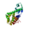 4lzmS C: citing same article ( S: Starting model for refinement |
|---|---|
| Similar structure data |


-Links
 PDBj
PDBj


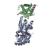
- Assembly
| Deposited unit | 
| ||||||||
|---|---|---|---|---|---|---|---|---|---|
| 1 | 
| ||||||||
| 2 | 
| ||||||||
| Unit cell |
| ||||||||
| Details | biological unit is the same as asym. |
-Components
| #1: Protein | Mass: 47858.023 Da / Num. of mol.: 2 Fragment: UNP P08483 residues 57-259, 482-563, UNP D9IEF7 residues 61-161 Source method: isolated from a genetically manipulated source Source: (gene. exp.) Enterobacteria phage T4 (virus)Gene: Chrm3, Chrm-3, e, T4Tp126 / Production host:   Spodoptera frugiperda (fall armyworm) / References: UniProt: P08483, UniProt: D9IEF7, lysozyme Spodoptera frugiperda (fall armyworm) / References: UniProt: P08483, UniProt: D9IEF7, lysozyme#2: Chemical |   Mass: 150.087 Da / Num. of mol.: 3 / Source method: obtained synthetically / Formula: C4H6O6 Mass: 150.087 Da / Num. of mol.: 3 / Source method: obtained synthetically / Formula: C4H6O6#3: Chemical | 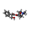  Mass: 318.387 Da / Num. of mol.: 2 / Source method: obtained synthetically / Formula: C18H24NO4 / Comment: medication, inhibitor*YM Mass: 318.387 Da / Num. of mol.: 2 / Source method: obtained synthetically / Formula: C18H24NO4 / Comment: medication, inhibitor*YMHas protein modification | Y | Sequence details | The fusion protein is a chimeric of M3 and RB69 lysozyme. The fusion protein is made of M 3 ( ...The fusion protein is a chimeric of M3 and RB69 lysozyme. The fusion protein is made of M 3 ( residues 57-259) - Lysozyme (residues 1000-1117)- M3 (residues 482-563). | |
|---|
-Experimental details
-Experiment
| Experiment | Method: X-RAY DIFFRACTION / Number of used crystals: 1 |
|---|
- Sample preparation
| Crystal | Density Matthews: 4.24 Å3/Da / Density % sol: 70.96 % |
|---|---|
| Crystal grow | Temperature: 293 K / Method: lipidic cubic phase Details: The best crystallization condition was 100 mM Tris pH 7.5, 44% PEG 300 and 400 mM ammonium tartrate. |
-Data collection
| Diffraction | Mean temperature: 80 K |
|---|---|
| Diffraction source | Source: SYNCHROTRON / Site: APS  / Beamline: 23-ID-D / Wavelength: 1.033 Å / Beamline: 23-ID-D / Wavelength: 1.033 Å |
| Detector | Type: MARMOSAIC 300 mm CCD / Detector: CCD / Date: Mar 16, 2013 |
| Radiation | Protocol: SINGLE WAVELENGTH / Monochromatic (M) / Laue (L): M / Scattering type: x-ray |
| Radiation wavelength | Wavelength: 1.033 Å / Relative weight: 1 |
| Reflection | Resolution: 3.7→33.7 Å / Num. obs: 14787 / % possible obs: 93.7 % / Redundancy: 2.6 % / Biso Wilson estimate: 119.14 Å2 / Rmerge(I) obs: 0.219 / Net I/σ(I): 8.9 |
| Reflection shell | Resolution: 3.7→3.83 Å / Redundancy: 2.5 % / Rmerge(I) obs: 0.884 / Mean I/σ(I) obs: 2.2 / % possible all: 89.1 |
- Processing
| Software |
| ||||||||||||||||||||||||||||||||||||||||||
|---|---|---|---|---|---|---|---|---|---|---|---|---|---|---|---|---|---|---|---|---|---|---|---|---|---|---|---|---|---|---|---|---|---|---|---|---|---|---|---|---|---|---|---|
| Refinement | Method to determine structure: MOLECULAR REPLACEMENT Starting model: 4DAJ, 4LZM Resolution: 3.7→33.67 Å / SU ML: 0.59 / Cross valid method: FREE R-VALUE / σ(F): 1.36 / Phase error: 34.09 / Stereochemistry target values: ML
| ||||||||||||||||||||||||||||||||||||||||||
| Solvent computation | Shrinkage radii: 0.9 Å / VDW probe radii: 1.11 Å / Solvent model: FLAT BULK SOLVENT MODEL | ||||||||||||||||||||||||||||||||||||||||||
| Refinement step | Cycle: LAST / Resolution: 3.7→33.67 Å
| ||||||||||||||||||||||||||||||||||||||||||
| Refine LS restraints |
| ||||||||||||||||||||||||||||||||||||||||||
| LS refinement shell |
| ||||||||||||||||||||||||||||||||||||||||||
| Refinement TLS params. | Method: refined / Origin x: -37.3762 Å / Origin y: -1.074 Å / Origin z: 5.547 Å
| ||||||||||||||||||||||||||||||||||||||||||
| Refinement TLS group | Selection details: all |
