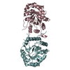
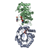
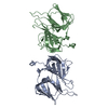
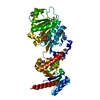
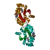
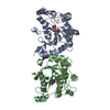
[English] 日本語
Yorodumi
- PDB-4qaf: Crystal structure of an engineered lipocalin (Anticalin) in compl... -

+
Open data


ID or keywords:

Loading...

-
Basic information

Entry
Database: PDB / ID: 4qaf
TitleCrystal structure of an engineered lipocalin (Anticalin) in complex with VEGF(8-109)
Components
  • Lipocalin-1
  • Vascular endothelial growth factor A
KeywordsTRANSPORT PROTEIN/SIGNALING PROTEIN / beta-barrel / binding protein / engineered lipocalin / TRANSPORT PROTEIN-SIGNALING PROTEIN complex
Function / homology
Function and homology information


Transport of fatty acids / basophil chemotaxis / positive regulation of endothelial cell chemotaxis by VEGF-activated vascular endothelial growth factor receptor signaling pathway / cellular stress response to acid chemical / VEGF-A complex / Signaling by VEGF / lymph vessel morphogenesis / positive regulation of lymphangiogenesis / negative regulation of adherens junction organization / vascular endothelial growth factor receptor 1 binding ...Transport of fatty acids / basophil chemotaxis / positive regulation of endothelial cell chemotaxis by VEGF-activated vascular endothelial growth factor receptor signaling pathway / cellular stress response to acid chemical / VEGF-A complex / Signaling by VEGF / lymph vessel morphogenesis / positive regulation of lymphangiogenesis / negative regulation of adherens junction organization / vascular endothelial growth factor receptor 1 binding / negative regulation of establishment of endothelial barrier / vascular endothelial growth factor receptor binding / VEGF ligand-receptor interactions / positive regulation of mast cell chemotaxis / post-embryonic camera-type eye development / primitive erythrocyte differentiation / positive regulation of protein kinase C signaling / positive regulation of cell proliferation by VEGF-activated platelet derived growth factor receptor signaling pathway / negative regulation of blood-brain barrier permeability / VEGF-activated neuropilin signaling pathway / bone trabecula formation / positive regulation of vascular endothelial growth factor signaling pathway / coronary vein morphogenesis / cardiac vascular smooth muscle cell development / lung vasculature development / lymphangiogenesis / eye photoreceptor cell development / endothelial cell chemotaxis / motor neuron migration / positive regulation of trophoblast cell migration / positive regulation of epithelial tube formation / vascular endothelial growth factor receptor-2 signaling pathway / VEGF binds to VEGFR leading to receptor dimerization / regulation of nitric oxide mediated signal transduction / positive regulation of axon extension involved in axon guidance / vascular wound healing / positive regulation of protein localization to early endosome / regulation of hematopoietic progenitor cell differentiation / positive regulation of blood vessel endothelial cell proliferation involved in sprouting angiogenesis / positive regulation of branching involved in ureteric bud morphogenesis / camera-type eye morphogenesis / neuropilin binding / induction of positive chemotaxis / coronary artery morphogenesis / negative regulation of cell-cell adhesion mediated by cadherin / vascular endothelial growth factor receptor 2 binding / tube formation / positive regulation of vascular permeability / dopaminergic neuron differentiation / commissural neuron axon guidance / negative regulation of epithelial to mesenchymal transition / platelet-derived growth factor receptor binding / surfactant homeostasis / response to stimulus / extracellular matrix binding / cell migration involved in sprouting angiogenesis / cardiac muscle cell development / epithelial cell maturation / sprouting angiogenesis / positive regulation of positive chemotaxis / endothelial cell proliferation / Regulation of gene expression by Hypoxia-inducible Factor / positive regulation of leukocyte migration / vascular endothelial growth factor signaling pathway / chloride ion binding / positive regulation of p38MAPK cascade / positive regulation of endothelial cell chemotaxis / artery morphogenesis / cysteine-type endopeptidase inhibitor activity / branching involved in blood vessel morphogenesis / positive regulation of cell migration involved in sprouting angiogenesis / positive regulation of DNA biosynthetic process / retinal ganglion cell axon guidance / positive regulation of neuroblast proliferation / positive chemotaxis / negative regulation of fat cell differentiation / transmembrane receptor protein tyrosine kinase activator activity / positive regulation of sprouting angiogenesis / chemoattractant activity / outflow tract morphogenesis / positive regulation of focal adhesion assembly / mesoderm development / monocyte differentiation / positive regulation of receptor internalization / macrophage differentiation / fibronectin binding / positive regulation of cell division / positive regulation of cell adhesion / neuroblast proliferation / positive regulation of blood vessel endothelial cell migration / cellular response to vascular endothelial growth factor stimulus / mammary gland alveolus development / vasculogenesis / positive regulation of osteoblast differentiation / vascular endothelial growth factor receptor signaling pathway / heart morphogenesis / ovarian follicle development / cell maturation / homeostasis of number of cells within a tissue / positive regulation of protein autophosphorylation
Similarity search - Function
von Ebner's gland protein/ Bos/Can allergen / Vascular endothelial growth factor, heparin-binding domain / Vascular endothelial growth factor, heparin-binding domain superfamily / VEGF heparin-binding domain / PDGF/VEGF domain / Platelet-derived growth factor, conserved site / PDGF/VEGF domain / Platelet-derived growth factor (PDGF) family signature. / Platelet-derived growth factor (PDGF) family profile. / Platelet-derived and vascular endothelial growth factors (PDGF, VEGF) family ...von Ebner's gland protein/ Bos/Can allergen / Vascular endothelial growth factor, heparin-binding domain / Vascular endothelial growth factor, heparin-binding domain superfamily / VEGF heparin-binding domain / PDGF/VEGF domain / Platelet-derived growth factor, conserved site / PDGF/VEGF domain / Platelet-derived growth factor (PDGF) family signature. / Platelet-derived growth factor (PDGF) family profile. / Platelet-derived and vascular endothelial growth factors (PDGF, VEGF) family / Cystine Knot Cytokines, subunit B / Cystine-knot cytokines / Lipocalin / Cystine-knot cytokine / Lipocalin / cytosolic fatty-acid binding protein family / Lipocalin/cytosolic fatty-acid binding domain / Calycin / Ribbon / Mainly Beta
Similarity search - Domain/homology
ACETATE ION / Chem-OMA / Vascular endothelial growth factor A, long form / Lipocalin-1
Similarity search - Component
Biological speciesHomo sapiens (human)
MethodX-RAY DIFFRACTION / SYNCHROTRON / SAD, MOLECULAR REPLACEMENT / Resolution: 1.8 Å
AuthorsGiese, T. / Skerra, A.
CitationJournal: To be Published
Title: Crystal structure of an Anticalin with specific blocking activity towards human vascular endothelial growth factor (VEGF) reveals plasticity of the lipocalin fold
Authors: Giese, T. / Skerra, A.
History
DepositionMay 4, 2014Deposition site: RCSB / Processing site: RCSB
Revision 1.0May 6, 2015Provider: repository / Type: Initial release
Revision 1.1Sep 20, 2023Group: Data collection / Database references ...Data collection / Database references / Derived calculations / Refinement description
Category: chem_comp_atom / chem_comp_bond ...chem_comp_atom / chem_comp_bond / database_2 / pdbx_initial_refinement_model / struct_ref_seq_dif / struct_site
Item: _database_2.pdbx_DOI / _database_2.pdbx_database_accession ..._database_2.pdbx_DOI / _database_2.pdbx_database_accession / _struct_ref_seq_dif.details / _struct_site.pdbx_auth_asym_id / _struct_site.pdbx_auth_comp_id / _struct_site.pdbx_auth_seq_id

-
Structure visualization

Structure viewerMolecule:
MolmilJmol/JSmol

Downloads & links

-
Assembly

Deposited unit
A: Lipocalin-1
C: Vascular endothelial growth factor A
D: Vascular endothelial growth factor A
B: Lipocalin-1
hetero molecules


Theoretical massNumber of molelcules
Total (without water)59,60310
Polymers58,6674
Non-polymers9366
Water3,783210
1


  • Idetical with deposited unit
  • defined by author&software
TypeNameSymmetry operationNumber
identity operation1_555x,y,z1
Buried area7340 Å2
ΔGint-104 kcal/mol
Surface area19670 Å2
MethodPISA
Unit cell
Length a, b, c (Å)88.210, 88.210, 103.421
Angle α, β, γ (deg.)90.000, 90.000, 120.000
Int Tables number154
Space group name H-MP3221

-
Components

-
Protein , 2 types, 4 molecules ABCD

#1: Protein Lipocalin-1 / Tear lipocalin / Tlc / Tear prealbumin / TP / Von Ebner gland protein / VEG protein


Mass: 16184.311 Da / Num. of mol.: 2 / Fragment: UNP residues 23-174
Mutation: R26V,E27G,F28A,P29L,E30R,M31C,N32L,L33A,E34G,T37I,M39T,L56H,S58K,C61S,E69S,K76I,D80I,K83I,Y87K,I89G,C101S,E104C,H106S,K108V,R111P,K114W,C153S
Source method: isolated from a genetically manipulated source
Source: (gene. exp.) Homo sapiens (human) / Gene: engineered variant, LCN1 / Plasmid: pTLc99 / Production host: Escherichia coli (E. coli) / Strain (production host): JM83 / References: UniProt: P31025
#2: Protein Vascular endothelial growth factor A / VEGF-A / Vascular permeability factor / VPF


Mass: 13148.979 Da / Num. of mol.: 2 / Fragment: UNP residues 34-135
Source method: isolated from a genetically manipulated source
Source: (gene. exp.) Homo sapiens (human) / Gene: VEGFA, VEGF / Plasmid: pVEGFs / Production host: Escherichia coli (E. coli) / Strain (production host): BL21(DE3) / References: UniProt: P15692

-
Non-polymers , 4 types, 216 molecules

#3: Chemical ChemComp-OMA / 10-{(1R,2R)-2-[(2E)-hex-2-en-1-yl]cyclopropyl}decanoic acid


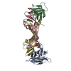
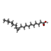
Mass: 294.472 Da / Num. of mol.: 2 / Source method: obtained synthetically / Formula: C19H34O2
#4: Chemical ChemComp-SO4 / SULFATE ION


Mass: 96.063 Da / Num. of mol.: 3 / Source method: obtained synthetically / Formula: SO4
#5: Chemical ChemComp-ACT / ACETATE ION


Mass: 59.044 Da / Num. of mol.: 1 / Source method: obtained synthetically / Formula: C2H3O2
#6: Water ChemComp-HOH / water


Mass: 18.015 Da / Num. of mol.: 210 / Source method: isolated from a natural source / Formula: H2O

-
Experimental details

-
Experiment

ExperimentMethod: X-RAY DIFFRACTION / Number of used crystals: 1

-
Sample preparation

CrystalDensity Matthews: 1.98 Å3/Da / Density % sol: 37.87 %
Crystal growTemperature: 293 K / Method: vapor diffusion, hanging drop / pH: 3.5
Details: 1 M sodium chloride, 0.2 M lithium sulfate, 7.5% w/v dextran sulfate, pH 3.5, VAPOR DIFFUSION, HANGING DROP, temperature 293K

-
Data collection

DiffractionMean temperature: 100 K
Diffraction sourceSource: SYNCHROTRON / Site: BESSY / Beamline: 14.1 / Wavelength: 0.91841 Å
DetectorType: DECTRIS PILATUS 6M / Detector: PIXEL / Date: May 29, 2009 / Details: mirrors
RadiationMonochromator: sagittally bent Si(111) / Protocol: SINGLE WAVELENGTH / Monochromatic (M) / Laue (L): M / Scattering type: x-ray
Radiation wavelengthWavelength: 0.91841 Å / Relative weight: 1
ReflectionResolution: 1.8→35 Å / Num. all: 43626 / Num. obs: 43539 / % possible obs: 99.8 % / Observed criterion σ(F): 0 / Observed criterion σ(I): -3 / Redundancy: 5.75 % / Biso Wilson estimate: 33.05 Å2 / Rmerge(I) obs: 0.041 / Net I/σ(I): 23.88
Reflection shell
Resolution (Å)Rmerge(I) obsMean I/σ(I) obsNum. measured obsNum. unique obsDiffraction-ID% possible all
1.8-1.850.4263.95185113189199.9
1.85-1.90.3055.44179133086199.9
1.9-1.950.2247.25175723027199.9
1.95-2.010.1898.641704729471100
2.01-2.080.14211.11652728361100
2.08-2.150.11913.231617927891100
2.15-2.230.09615.98154232631199.9
2.23-2.320.07619.14148902578199.7
2.32-2.430.06521.76144572471199.9
2.43-2.550.05824.52136042342199.9
2.55-2.680.04629.491308322581100
2.68-2.850.04233.531235421371100
2.85-3.040.03737.89116622012199.9
3.04-3.290.03144.44107161870199.9
3.29-3.60.02950.197801723199.8
3.6-4.020.02852.7289271593199.8
4.02-4.650.02656.5577901398199.8
4.65-5.690.02556.5466371194199.9
5.69-8.050.02455.615176953199.7
8.05-350.02848.482217505191

-
Processing

Software
NameVersionClassificationNB
XSCALEdata scaling
REFMACrefinement
PDB_EXTRACT3.11data extraction
MAR345dtbdata collection
XDSdata reduction
Auto-Rickshawphasing
RefinementMethod to determine structure: SAD, MOLECULAR REPLACEMENT
Starting model: PDB ENTRY 1BJ1
Resolution: 1.8→34.47 Å / Cor.coef. Fo:Fc: 0.941 / Cor.coef. Fo:Fc free: 0.934 / WRfactor Rfree: 0.2579 / WRfactor Rwork: 0.215 / Occupancy max: 1 / Occupancy min: 0 / FOM work R set: 0.8466 / SU B: 2.565 / SU ML: 0.081 / SU R Cruickshank DPI: 0.1282 / SU Rfree: 0.1258 / Cross valid method: THROUGHOUT / σ(F): 0 / ESU R: 0.128 / ESU R Free: 0.126 / Stereochemistry target values: MAXIMUM LIKELIHOOD / Details: HYDROGENS HAVE BEEN USED IF PRESENT IN THE INPUT
RfactorNum. reflection% reflectionSelection details
Rfree0.2421 2195 5 %RANDOM
Rwork0.2033 ---
obs0.2053 41384 99.91 %-
all-43626 --
Solvent computationIon probe radii: 0.8 Å / Shrinkage radii: 0.8 Å / VDW probe radii: 1.2 Å / Solvent model: MASK
Displacement parametersBiso max: 114.42 Å2 / Biso mean: 33.7489 Å2 / Biso min: 9.38 Å2
Baniso -1Baniso -2Baniso -3
1-0 Å20 Å20 Å2
2--0 Å20 Å2
3----0 Å2
Refinement stepCycle: LAST / Resolution: 1.8→34.47 Å
ProteinNucleic acidLigandSolventTotal
Num. atoms3029 0 61 210 3300
Refine LS restraints
Refine-IDTypeDev idealDev ideal targetNumber
X-RAY DIFFRACTIONr_bond_refined_d0.0210.023191
X-RAY DIFFRACTIONr_angle_refined_deg2.2141.984323
X-RAY DIFFRACTIONr_dihedral_angle_1_deg7.1315392
X-RAY DIFFRACTIONr_dihedral_angle_2_deg37.33324.58131
X-RAY DIFFRACTIONr_dihedral_angle_3_deg17.05715550
X-RAY DIFFRACTIONr_dihedral_angle_4_deg11.9811513
X-RAY DIFFRACTIONr_chiral_restr0.1650.2484
X-RAY DIFFRACTIONr_gen_planes_refined0.0120.0212341
LS refinement shellResolution: 1.8→1.847 Å / Total num. of bins used: 20
RfactorNum. reflection% reflection
Rfree0.366 152 -
Rwork0.31 2839 -
all-2991 -
obs--100 %

+
About Yorodumi

-
News

-
Feb 9, 2022. New format data for meta-information of EMDB entries

New format data for meta-information of EMDB entries

  • Version 3 of the EMDB header file is now the official format.
  • The previous official version 1.9 will be removed from the archive.

Related info.:EMDB header

External links:wwPDB to switch to version 3 of the EMDB data model

-
Aug 12, 2020. Covid-19 info

Covid-19 info

URL: https://pdbjlvh1.pdbj.org/emnavi/covid19.php

New page: Covid-19 featured information page in EM Navigator.

Related info.:Covid-19 info / Mar 5, 2020. Novel coronavirus structure data

+
Mar 5, 2020. Novel coronavirus structure data

Novel coronavirus structure data

Related info.:Yorodumi Speices / Aug 12, 2020. Covid-19 info

External links:COVID-19 featured content - PDBj / Molecule of the Month (242):Coronavirus Proteases

+
Jan 31, 2019. EMDB accession codes are about to change! (news from PDBe EMDB page)

EMDB accession codes are about to change! (news from PDBe EMDB page)

  • The allocation of 4 digits for EMDB accession codes will soon come to an end. Whilst these codes will remain in use, new EMDB accession codes will include an additional digit and will expand incrementally as the available range of codes is exhausted. The current 4-digit format prefixed with “EMD-” (i.e. EMD-XXXX) will advance to a 5-digit format (i.e. EMD-XXXXX), and so on. It is currently estimated that the 4-digit codes will be depleted around Spring 2019, at which point the 5-digit format will come into force.
  • The EM Navigator/Yorodumi systems omit the EMD- prefix.

Related info.:Q: What is EMD? / ID/Accession-code notation in Yorodumi/EM Navigator

External links:EMDB Accession Codes are Changing Soon! / Contact to PDBj

+
Jul 12, 2017. Major update of PDB

Major update of PDB

  • wwPDB released updated PDB data conforming to the new PDBx/mmCIF dictionary.
  • This is a major update changing the version number from 4 to 5, and with Remediation, in which all the entries are updated.
  • In this update, many items about electron microscopy experimental information are reorganized (e.g. em_software).
  • Now, EM Navigator and Yorodumi are based on the updated data.

External links:wwPDB Remediation / Enriched Model Files Conforming to OneDep Data Standards Now Available in the PDB FTP Archive

-
Yorodumi

Thousand views of thousand structures

  • Yorodumi is a browser for structure data from EMDB, PDB, SASBDB, etc.
  • This page is also the successor to EM Navigator detail page, and also detail information page/front-end page for Omokage search.
  • The word "yorodu" (or yorozu) is an old Japanese word meaning "ten thousand". "mi" (miru) is to see.

Related info.:EMDB / PDB / SASBDB / Comparison of 3 databanks / Yorodumi Search / Aug 31, 2016. New EM Navigator & Yorodumi / Yorodumi Papers / Jmol/JSmol / Function and homology information / Changes in new EM Navigator and Yorodumi

Read more