Mass: 392.512 Da / Num. of mol.: 1 / Source method: obtained synthetically / Formula: C19H22NO4S2 / Comment: antagonist*YM
Has protein modification
Y
Sequence details
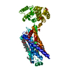
The fusion protein is a chimeric of M3 and T4: T4L lysozyme was inserted into a intracellular loop ...The fusion protein is a chimeric of M3 and T4: T4L lysozyme was inserted into a intracellular loop of the receptor. The fusion protein is made of M 3 ( residues 57-259) - T4L (residues 1001-1161)- M3 (residues 482-563). These two proteins were numbered separately in order to maintain original (UNP P08483 and P00720) numbering.
-
Experimental details
-
Experiment
Experiment
Method: X-RAY DIFFRACTION / Number of used crystals: 1
-
Sample preparation
Crystal
Density Matthews: 2.49 Å3/Da / Density % sol: 50.55 %
Crystal grow
Temperature: 298 K / Method: lipidic cubic phase / pH: 8.1 Details: 45% PEG 300, 110 mM ammonium sulfate, 113.5 mM lithium citrate, 100 mM Tris
In the structure databanks used in Yorodumi, some data are registered as the other names, "COVID-19 virus" and "2019-nCoV". Here are the details of the virus and the list of structure data.
Jan 31, 2019. EMDB accession codes are about to change! (news from PDBe EMDB page)
EMDB accession codes are about to change! (news from PDBe EMDB page)
The allocation of 4 digits for EMDB accession codes will soon come to an end. Whilst these codes will remain in use, new EMDB accession codes will include an additional digit and will expand incrementally as the available range of codes is exhausted. The current 4-digit format prefixed with “EMD-” (i.e. EMD-XXXX) will advance to a 5-digit format (i.e. EMD-XXXXX), and so on. It is currently estimated that the 4-digit codes will be depleted around Spring 2019, at which point the 5-digit format will come into force.
The EM Navigator/Yorodumi systems omit the EMD- prefix.
Related info.:Q: What is EMD? / ID/Accession-code notation in Yorodumi/EM Navigator
Yorodumi is a browser for structure data from EMDB, PDB, SASBDB, etc.
This page is also the successor to EM Navigator detail page, and also detail information page/front-end page for Omokage search.
The word "yorodu" (or yorozu) is an old Japanese word meaning "ten thousand". "mi" (miru) is to see.
Related info.:EMDB / PDB / SASBDB / Comparison of 3 databanks / Yorodumi Search / Aug 31, 2016. New EM Navigator & Yorodumi / Yorodumi Papers / Jmol/JSmol / Function and homology information / Changes in new EM Navigator and Yorodumi
 Movie
Movie Controller

 Yorodumi
 Open data
 Basic information
 Components
 Keywords
 Function and homology information
 Enterobacteria phage T4 (virus)
 X-RAY DIFFRACTION /
  Authors
 United States,
  Denmark, 4items
  Citation
 Structure visualization
 Downloads & links
 Other downloads


 PDBj
PDBj


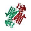
 Assembly
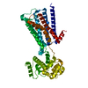

 Spodoptera frugiperda (fall armyworm) / References: UniProt: P08483, UniProt: P00720, lysozyme
 Mass: 392.512 Da / Num. of mol.: 1 / Source method: obtained synthetically / Formula: C19H22NO4S2 / Comment: antagonist*YM
 Sample preparation
 Processing