 Movie
Movie Controller


+ Open data

- Basic information
| Entry | Database: PDB / ID: 4tsy | ||||||
|---|---|---|---|---|---|---|---|
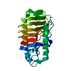
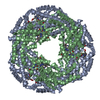
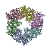
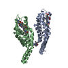
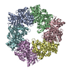
| Title | Crystal structure of FraC with lipids | ||||||
 Components Components | Fragaceatoxin C | ||||||
 Keywords Keywords | TOXIN / actinoporin pore-forming toxin / Membrane lipids / Phosphocholine / Lipid-protein interaction | ||||||
| Function / homology |  Function and homology information Function and homology informationnematocyst / cytolysis in another organism / pore complex assembly / other organism cell membrane / pore complex / monoatomic cation transport / toxin activity / channel activity / lipid binding / extracellular region / identical protein binding Similarity search - Function | ||||||
| Biological species |  | ||||||
| Method |  X-RAY DIFFRACTION / SYNCHROTRON / MOLECULAR REPLACEMENT / Resolution: 3.14 Å X-RAY DIFFRACTION / SYNCHROTRON / MOLECULAR REPLACEMENT / Resolution: 3.14 Å | ||||||
 Authors Authors | Caaveiro, J.M.M. / Tanaka, K. / Tsumoto, K. | ||||||
 Citation Citation | Journal: Nat Commun / Year: 2015 Title: Structural basis for self-assembly of a cytolytic pore lined by protein and lipid Authors: Tanaka, K. / Caaveiro, J.M.M. / Morante, K. / Gonzalez-Manas, J.M. / Tsumoto, K. #1: Journal: Toxicon / Year: 2009 Title: Purification, cloning and characterization of fragaceatoxin C, a novel actinoporin from the sea anemone Actinia fragacea Authors: Bellomio, A. / Morante, K. / Barlic, A. / Gutierrez-Aguirre, I. / Viguera, A.R. / Gonzalez-Manas, J.M. | ||||||
| History |
|
- Structure visualization
| Structure viewer | Molecule: MolmilJmol/JSmol |
|---|
- Downloads & links
-Download
| PDBx/mmCIF format | 4tsy.cif.gz | 308 KB | Display | PDBx/mmCIF format |
|---|---|---|---|---|
| PDB format | pdb4tsy.ent.gz | 256.3 KB | Display | PDB format |
| PDBx/mmJSON format | 4tsy.json.gz | Tree view | PDBx/mmJSON format | |
| Others |  Other downloads Other downloads |
-Validation report
| Arichive directory | https://data.pdbj.org/pub/pdb/validation_reports/ts/4tsyftp://data.pdbj.org/pub/pdb/validation_reports/ts/4tsy | HTTPS FTP |
|---|
-Related structure data
| Related structure data | 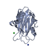 3vwiSC  3w9pC 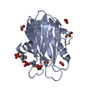 4tslC  4tsnC  4tsoC 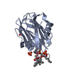 4tspC 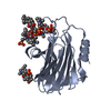 4tsqC S: Starting model for refinement C: citing same article ( |
|---|---|
| Similar structure data |


-Links
 PDBj
PDBj
- Assembly
| Deposited unit | 
| ||||||||||||||||||||||||||||||||||||||||||||||||||||||||||||||||||||||||||||||||||||||||||||||||||
|---|---|---|---|---|---|---|---|---|---|---|---|---|---|---|---|---|---|---|---|---|---|---|---|---|---|---|---|---|---|---|---|---|---|---|---|---|---|---|---|---|---|---|---|---|---|---|---|---|---|---|---|---|---|---|---|---|---|---|---|---|---|---|---|---|---|---|---|---|---|---|---|---|---|---|---|---|---|---|---|---|---|---|---|---|---|---|---|---|---|---|---|---|---|---|---|---|---|---|---|
| 1 | 
| ||||||||||||||||||||||||||||||||||||||||||||||||||||||||||||||||||||||||||||||||||||||||||||||||||
| Unit cell |
| ||||||||||||||||||||||||||||||||||||||||||||||||||||||||||||||||||||||||||||||||||||||||||||||||||
| Noncrystallographic symmetry (NCS) | NCS domain:
NCS domain segments: Component-ID: _ / Beg auth comp-ID: VAL / Beg label comp-ID: VAL / End auth comp-ID: ALA / End label comp-ID: ALA / Refine code: _ / Auth seq-ID: 4 - 179 / Label seq-ID: 4 - 179
NCS ensembles :
|
-Components
| #1: Protein | Mass: 19746.303 Da / Num. of mol.: 4 Source method: isolated from a genetically manipulated source Source: (gene. exp.)  #2: Chemical | ChemComp-HXJ /   Mass: 451.558 Da / Num. of mol.: 12 / Source method: obtained synthetically / Formula: C21H44N2O6P Mass: 451.558 Da / Num. of mol.: 12 / Source method: obtained synthetically / Formula: C21H44N2O6P#3: Chemical | ChemComp-SO4 /   Mass: 96.063 Da / Num. of mol.: 19 / Source method: obtained synthetically / Formula: SO4 Mass: 96.063 Da / Num. of mol.: 19 / Source method: obtained synthetically / Formula: SO4#4: Chemical | ChemComp-HTO / |   Mass: 148.200 Da / Num. of mol.: 1 / Source method: obtained synthetically / Formula: C7H16O3 Mass: 148.200 Da / Num. of mol.: 1 / Source method: obtained synthetically / Formula: C7H16O3#5: Water | ChemComp-HOH / |  Mass: 18.015 Da / Num. of mol.: 17 / Source method: isolated from a natural source / Formula: H2O Mass: 18.015 Da / Num. of mol.: 17 / Source method: isolated from a natural source / Formula: H2O |
|---|
-Experimental details
-Experiment
| Experiment | Method: X-RAY DIFFRACTION |
|---|
- Sample preparation
| Crystal | Density Matthews: 5.4 Å3/Da / Density % sol: 77.07 % |
|---|---|
| Crystal grow | Temperature: 293 K / Method: lipidic cubic phase / pH: 7 Details: Jeffamine M600, 1,2,3-heptanetriol, monoolein, ammonium sulfate, HEPES |
-Data collection
| Diffraction | Mean temperature: 100 K |
|---|---|
| Diffraction source | Source: SYNCHROTRON / Site: Photon Factory  / Beamline: AR-NE3A / Wavelength: 1 Å / Beamline: AR-NE3A / Wavelength: 1 Å |
| Detector | Type: ADSC QUANTUM 270 / Detector: CCD / Date: Feb 17, 2014 |
| Radiation | Protocol: SINGLE WAVELENGTH / Monochromatic (M) / Laue (L): M / Scattering type: x-ray |
| Radiation wavelength | Wavelength: 1 Å / Relative weight: 1 |
| Reflection | Resolution: 3.14→38.65 Å / Num. obs: 30154 / % possible obs: 94.6 % / Redundancy: 7.3 % / Biso Wilson estimate: 42.4 Å2 / Rmerge(I) obs: 0.15 / Net I/σ(I): 9.9 |
| Reflection shell | Resolution: 3.14→3.32 Å / Redundancy: 5.5 % / Rmerge(I) obs: 0.41 / Mean I/σ(I) obs: 3.4 / % possible all: 83.5 |
- Processing
| Software | Name: REFMAC / Version: 5.8.0049 / Classification: refinement | ||||||||||||||||||||||||||||||||||||||||||||||||||||||||||||||||||||||||||||||||||||||||||||||||||||||||||||||||||||||||||||||||||||||||||||||||||||||||||||||||||||||||||||||||||||||
|---|---|---|---|---|---|---|---|---|---|---|---|---|---|---|---|---|---|---|---|---|---|---|---|---|---|---|---|---|---|---|---|---|---|---|---|---|---|---|---|---|---|---|---|---|---|---|---|---|---|---|---|---|---|---|---|---|---|---|---|---|---|---|---|---|---|---|---|---|---|---|---|---|---|---|---|---|---|---|---|---|---|---|---|---|---|---|---|---|---|---|---|---|---|---|---|---|---|---|---|---|---|---|---|---|---|---|---|---|---|---|---|---|---|---|---|---|---|---|---|---|---|---|---|---|---|---|---|---|---|---|---|---|---|---|---|---|---|---|---|---|---|---|---|---|---|---|---|---|---|---|---|---|---|---|---|---|---|---|---|---|---|---|---|---|---|---|---|---|---|---|---|---|---|---|---|---|---|---|---|---|---|---|---|
| Refinement | Method to determine structure: MOLECULAR REPLACEMENT Starting model: 3VWI Resolution: 3.14→38.65 Å / Cor.coef. Fo:Fc: 0.919 / Cor.coef. Fo:Fc free: 0.889 / SU B: 33.743 / SU ML: 0.258 / Cross valid method: THROUGHOUT / ESU R: 0.732 / ESU R Free: 0.331 / Stereochemistry target values: MAXIMUM LIKELIHOOD / Details: HYDROGENS HAVE BEEN ADDED IN THE RIDING POSITIONS
| ||||||||||||||||||||||||||||||||||||||||||||||||||||||||||||||||||||||||||||||||||||||||||||||||||||||||||||||||||||||||||||||||||||||||||||||||||||||||||||||||||||||||||||||||||||||
| Solvent computation | Ion probe radii: 0.8 Å / Shrinkage radii: 0.8 Å / VDW probe radii: 1.2 Å / Solvent model: MASK | ||||||||||||||||||||||||||||||||||||||||||||||||||||||||||||||||||||||||||||||||||||||||||||||||||||||||||||||||||||||||||||||||||||||||||||||||||||||||||||||||||||||||||||||||||||||
| Displacement parameters | Biso mean: 52.754 Å2
| ||||||||||||||||||||||||||||||||||||||||||||||||||||||||||||||||||||||||||||||||||||||||||||||||||||||||||||||||||||||||||||||||||||||||||||||||||||||||||||||||||||||||||||||||||||||
| Refinement step | Cycle: 1 / Resolution: 3.14→38.65 Å
| ||||||||||||||||||||||||||||||||||||||||||||||||||||||||||||||||||||||||||||||||||||||||||||||||||||||||||||||||||||||||||||||||||||||||||||||||||||||||||||||||||||||||||||||||||||||
| Refine LS restraints |
|