 Movie
Movie Controller

[English] 日本語
 Yorodumi
- PDB-4s3l: Crystal Structure of major pilin protein PitB from type II pilus ... -
+ Open data

- Basic information
| Entry | Database: PDB / ID: 4s3l | ||||||
|---|---|---|---|---|---|---|---|
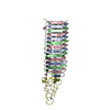
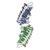
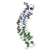
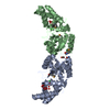
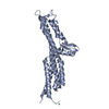
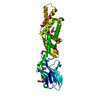
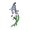
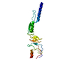
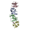
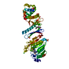
| Title | Crystal Structure of major pilin protein PitB from type II pilus of Streptococcus pneumoniae | ||||||
 Components Components | Major Pilin Protein | ||||||
 Keywords Keywords | CELL ADHESION / Major pilin / Type II pilus / virulence / CNAB / IgG fold / IgG-rev fold / Pilin protein | ||||||
| Function / homology | Spy0128-like isopeptide containing domain / Streptococcal pilin isopeptide linker superfamily / : / Domain of unknown function (DUF7601) / Domain of unknown function (DUF5979) / membrane / PitB Function and homology information Function and homology information | ||||||
| Biological species |   Streptococcus pneumoniae (bacteria) Streptococcus pneumoniae (bacteria) | ||||||
| Method |  X-RAY DIFFRACTION / SYNCHROTRON / SAD / Resolution: 2.8 Å X-RAY DIFFRACTION / SYNCHROTRON / SAD / Resolution: 2.8 Å | ||||||
 Authors Authors | Shaik, M.M. / Dessen, A. / Di Guilmi, A.M. | ||||||
 Citation Citation | Journal: J.Biol.Chem. / Year: 2015 Title: A Structural Snapshot of Type II Pilus Formation in Streptococcus pneumoniae. Authors: Shaik, M.M. / Lombardi, C. / Maragno Trindade, D. / Fenel, D. / Schoehn, G. / Di Guilmi, A.M. / Dessen, A. | ||||||
| History |
|
- Structure visualization
| Structure viewer | Molecule: MolmilJmol/JSmol |
|---|
- Downloads & links
-Download
| PDBx/mmCIF format | 4s3l.cif.gz | 140.5 KB | Display | PDBx/mmCIF format |
|---|---|---|---|---|
| PDB format | pdb4s3l.ent.gz | 110.6 KB | Display | PDB format |
| PDBx/mmJSON format | 4s3l.json.gz | Tree view | PDBx/mmJSON format | |
| Others |  Other downloads Other downloads |
-Validation report
| Arichive directory | https://data.pdbj.org/pub/pdb/validation_reports/s3/4s3lftp://data.pdbj.org/pub/pdb/validation_reports/s3/4s3l | HTTPS FTP |
|---|
-Related structure data

-Links
 PDBj
PDBj- Assembly
| Deposited unit | 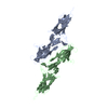
| ||||||||
|---|---|---|---|---|---|---|---|---|---|
| 1 | 
| ||||||||
| 2 | 
| ||||||||
| Unit cell |
|
-Components
| #1: Protein | Mass: 37197.891 Da / Num. of mol.: 2 / Fragment: UNP residues 46-386 Source method: isolated from a genetically manipulated source Source: (gene. exp.) Streptococcus pneumoniae (strain Taiwan19F-14) (bacteria)Strain: Taiwan19F-14 / Gene: pitB / Plasmid: pet151 / Production host: #2: Water | ChemComp-HOH / |  Mass: 18.015 Da / Num. of mol.: 79 / Source method: isolated from a natural source / Formula: H2O Mass: 18.015 Da / Num. of mol.: 79 / Source method: isolated from a natural source / Formula: H2OHas protein modification | Y | |
|---|
-Experimental details
-Experiment
| Experiment | Method: X-RAY DIFFRACTION / Number of used crystals: 1 |
|---|
- Sample preparation
| Crystal | Density Matthews: 3.57 Å3/Da / Density % sol: 65.54 % |
|---|---|
| Crystal grow | Temperature: 293 K / Method: vapor diffusion / pH: 7 Details: 3M NaCl, 0.1M HEPES, pH 7.0, VAPOR DIFFUSION, temperature 293K |
-Data collection
| Diffraction |
| ||||||||||||||||||
|---|---|---|---|---|---|---|---|---|---|---|---|---|---|---|---|---|---|---|---|
| Diffraction source |
| ||||||||||||||||||
| Detector |
| ||||||||||||||||||
| Radiation |
| ||||||||||||||||||
| Radiation wavelength | Wavelength: 0.939 Å / Relative weight: 1 | ||||||||||||||||||
| Reflection | Resolution: 2.8→54.21 Å / Num. obs: 25595 / % possible obs: 80 % / Observed criterion σ(F): 2.1 / Observed criterion σ(I): 2 / Redundancy: 4.8 % / Rmerge(I) obs: 0.195 / Net I/σ(I): 5.9 | ||||||||||||||||||
| Reflection shell | Resolution: 2.8→2.95 Å / Redundancy: 4.9 % / Rmerge(I) obs: 0.583 / Mean I/σ(I) obs: 2.1 / % possible all: 100 |

- Processing
| Software |
| ||||||||||||||||||||||||||||||||||||||||||||||||||||||||||||||||||||||
|---|---|---|---|---|---|---|---|---|---|---|---|---|---|---|---|---|---|---|---|---|---|---|---|---|---|---|---|---|---|---|---|---|---|---|---|---|---|---|---|---|---|---|---|---|---|---|---|---|---|---|---|---|---|---|---|---|---|---|---|---|---|---|---|---|---|---|---|---|---|---|---|
| Refinement | Method to determine structure: SAD / Resolution: 2.8→54.21 Å / SU ML: 0.58 / σ(F): 1.35 / Phase error: 39.87 / Stereochemistry target values: ML
| ||||||||||||||||||||||||||||||||||||||||||||||||||||||||||||||||||||||
| Solvent computation | Shrinkage radii: 0.9 Å / VDW probe radii: 1.11 Å / Solvent model: FLAT BULK SOLVENT MODEL | ||||||||||||||||||||||||||||||||||||||||||||||||||||||||||||||||||||||
| Refinement step | Cycle: LAST / Resolution: 2.8→54.21 Å
| ||||||||||||||||||||||||||||||||||||||||||||||||||||||||||||||||||||||
| Refine LS restraints |
| ||||||||||||||||||||||||||||||||||||||||||||||||||||||||||||||||||||||
| LS refinement shell |
|
