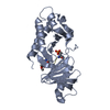
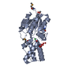
- PDB-2hsz: Crystal structure of a predicted phosphoglycolate phosphatase (hs... -
+
Open data
ID or keywords:
Loading...
-
Basic information
Entry
Database: PDB / ID: 2hsz
Title
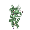
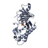
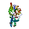
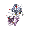
Crystal structure of a predicted phosphoglycolate phosphatase (hs_0176) from haemophilus somnus 129pt at 1.90 A resolution
Components
novel predicted phosphatase
Keywords
HYDROLASE / Structural genomics / Joint Center for Structural Genomics / JCSG / Protein Structure Initiative / PSI-2
Function / homology
Function and homology information
phosphoglycolate phosphatase / phosphoglycolate phosphatase activity / glycolate biosynthetic process / carbohydrate metabolic process / DNA repair / metal ion binding / cytosol Similarity search - Function
Phosphoglycolate phosphatase, prokaryotic / Hypothetical cof family signature 1. / : / Haloacid dehalogenase-like hydrolase / Haloacid dehalogenase-like hydrolase / Putative phosphatase; domain 2 / Phosphoglycolate phosphatase-like, domain 2 / HAD hydrolase, subfamily IA / HAD superfamily/HAD-like / HAD superfamily ...Phosphoglycolate phosphatase, prokaryotic / Hypothetical cof family signature 1. / : / Haloacid dehalogenase-like hydrolase / Haloacid dehalogenase-like hydrolase / Putative phosphatase; domain 2 / Phosphoglycolate phosphatase-like, domain 2 / HAD hydrolase, subfamily IA / HAD superfamily/HAD-like / HAD superfamily / HAD-like superfamily / DNA polymerase; domain 1 / Rossmann fold / Orthogonal Bundle / 3-Layer(aba) Sandwich / Mainly Alpha / Alpha Beta Similarity search - Domain/homology
REMARK 300 BIOMOLECULE: 1,2 THIS ENTRY CONTAINS THE CRYSTALLOGRAPHIC ASYMMETRIC UNIT WHICH CONSISTS ...REMARK 300 BIOMOLECULE: 1,2 THIS ENTRY CONTAINS THE CRYSTALLOGRAPHIC ASYMMETRIC UNIT WHICH CONSISTS OF 2 CHAIN(S). SEE REMARK 350 FOR INFORMATION ON GENERATING THE BIOLOGICAL MOLECULE(S). SIZE EXCLUSION CHROMATOGRAPHY DATA SUPPORTS THE ASSIGNMENT OF A MONOMER AS A BIOLOGICALLY SIGNIFICANT OLIGOMERIZATION STATE.
Remark 999
SEQUENCE A suitable database reference was not available at the time of processing.
Resolution: 1.897→29.988 Å / Num. obs: 51412 / % possible obs: 100 % / Redundancy: 4.8 % / Rmerge(I) obs: 0.074 / Rsym value: 0.074 / Net I/σ(I): 6.9
Reflection shell
Diffraction-ID: 1
Resolution (Å)
Redundancy (%)
Rmerge(I) obs
Mean I/σ(I) obs
Num. measured all
Num. unique all
Rsym value
% possible all
1.9-1.95
3.7
0.653
1
13790
3744
0.653
100
1.95-2
3.7
0.495
1.4
13483
3653
0.495
100
2-2.06
3.7
0.387
1.9
13114
3552
0.387
100
2.06-2.12
3.7
0.31
2.1
12717
3447
0.31
100
2.12-2.19
3.7
0.245
2.9
12562
3391
0.245
100
2.19-2.27
3.7
0.2
3.6
11886
3220
0.2
100
2.27-2.36
3.7
0.173
4.3
11632
3144
0.173
100
2.36-2.45
3.7
0.139
5.2
11223
3027
0.139
100
2.45-2.56
3.7
0.117
6.3
10840
2930
0.117
100
2.56-2.69
3.7
0.097
7.6
10205
2768
0.097
100
2.69-2.83
3.7
0.079
9
9840
2663
0.079
100
2.83-3
4.9
0.097
6.5
12359
2514
0.097
100
3-3.21
7.8
0.1
6.6
18390
2363
0.1
100
3.21-3.47
7.8
0.079
8.1
17250
2225
0.079
100
3.47-3.8
7.7
0.063
9.7
15749
2036
0.063
100
3.8-4.25
7.7
0.053
12.1
14441
1883
0.053
100
4.25-4.91
7.6
0.053
11.5
12581
1652
0.053
100
4.91-6.01
7.5
0.06
9.1
10679
1424
0.06
100
6.01-8.5
7.3
0.055
10.2
8281
1127
0.055
99.9
8.5-29.99
6.8
0.05
9.7
4406
649
0.05
97.6
-
Phasing
Phasing
Method: MAD
-
Processing
Software
Name
Version
Classification
NB
MolProbity
3beta29
modelbuilding
SHELX
phasing
REFMAC
5.2.0019
refinement
SCALA
datascaling
PDB_EXTRACT
2
dataextraction
MOSFLM
datareduction
CCP4
(SCALA)
datascaling
autoSHARP
phasing
Refinement
Method to determine structure: MAD / Resolution: 1.9→29.988 Å / Cor.coef. Fo:Fc: 0.963 / Cor.coef. Fo:Fc free: 0.953 / SU B: 5.066 / SU ML: 0.076 / TLS residual ADP flag: LIKELY RESIDUAL / Cross valid method: THROUGHOUT / σ(F): 0 / ESU R: 0.113 / ESU R Free: 0.112 Stereochemistry target values: MAXIMUM LIKELIHOOD WITH PHASES Details: (1). HYDROGENS HAVE BEEN ADDED IN THE RIDING POSITIONS. (2). A MET-INHIBITION PROTOCOL WAS USED FOR SELENOMETHIONINE INCORPORATION DURING PROTEIN EXPRESSION. THE OCCUPANCY OF THE SE ATOMS IN ...Details: (1). HYDROGENS HAVE BEEN ADDED IN THE RIDING POSITIONS. (2). A MET-INHIBITION PROTOCOL WAS USED FOR SELENOMETHIONINE INCORPORATION DURING PROTEIN EXPRESSION. THE OCCUPANCY OF THE SE ATOMS IN THE MSE RESIDUES WAS REDUCED TO 0.75 TO ACCOUNT FOR THE REDUCED SCATTERING POWER DUE TO PARTIAL S-MET INCORPORATION. (3). THERE ARE UNKNOWN DENSITY NEAR ACTIVE SITE IN A AND B CHAINS. THEY ARE NAMED AS UNL. (4). THERE ARE UNKNOWN DENSITY NEAR PHE 75 IN A AND B MOLECULES, TYR 77 AND PHE 138 IN A MOLECULE, PRO 152 AND TYR 200 IN B MOLECULE. THEY ARE ALL IN THE SOLVENT REGION AND LEFT UNMODELED. (5). ATOM RECORDS CONTAIN RESIDUAL B FACTORS ONLY.
Rfactor
Num. reflection
% reflection
Selection details
Rfree
0.205
2613
5.1 %
RANDOM
Rwork
0.169
-
-
-
obs
0.171
51362
99.75 %
-
Solvent computation
Ion probe radii: 0.8 Å / Shrinkage radii: 0.8 Å / VDW probe radii: 1.2 Å / Solvent model: MASK
In the structure databanks used in Yorodumi, some data are registered as the other names, "COVID-19 virus" and "2019-nCoV". Here are the details of the virus and the list of structure data.
Jan 31, 2019. EMDB accession codes are about to change! (news from PDBe EMDB page)
EMDB accession codes are about to change! (news from PDBe EMDB page)
The allocation of 4 digits for EMDB accession codes will soon come to an end. Whilst these codes will remain in use, new EMDB accession codes will include an additional digit and will expand incrementally as the available range of codes is exhausted. The current 4-digit format prefixed with “EMD-” (i.e. EMD-XXXX) will advance to a 5-digit format (i.e. EMD-XXXXX), and so on. It is currently estimated that the 4-digit codes will be depleted around Spring 2019, at which point the 5-digit format will come into force.
The EM Navigator/Yorodumi systems omit the EMD- prefix.
Related info.:Q: What is EMD? / ID/Accession-code notation in Yorodumi/EM Navigator
Yorodumi is a browser for structure data from EMDB, PDB, SASBDB, etc.
This page is also the successor to EM Navigator detail page, and also detail information page/front-end page for Omokage search.
The word "yorodu" (or yorozu) is an old Japanese word meaning "ten thousand". "mi" (miru) is to see.
Related info.:EMDB / PDB / SASBDB / Comparison of 3 databanks / Yorodumi Search / Aug 31, 2016. New EM Navigator & Yorodumi / Yorodumi Papers / Jmol/JSmol / Function and homology information / Changes in new EM Navigator and Yorodumi
 Movie
Movie Controller

 Yorodumi
 Open data
 Basic information
 Components
 Keywords
 Function and homology information
 Haemophilus somnus 129PT (bacteria)
 X-RAY DIFFRACTION /
  Authors
 Citation
 Structure visualization
 Downloads & links
 Other downloads


 PDBj
PDBj


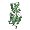
 Assembly
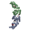

 Mass: 59.044 Da / Num. of mol.: 2 / Source method: obtained synthetically / Formula: C2H3O2
 Mass: 35.453 Da / Num. of mol.: 2 / Source method: obtained synthetically / Formula: Cl
 Mass: 18.015 Da / Num. of mol.: 334 / Source method: isolated from a natural source / Formula: H2O
 Sample preparation
 / Beamline: BL9-2 / Wavelength: 0.91837,0.97925,1.28281,0.97914
 Processing