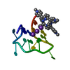
regulation of protein deubiquitination / negative regulation of cytoplasmic pattern recognition receptor signaling pathway / ubiquitin-like protein transferase activity / cellular response to tumor cell / nucleotide-binding domain, leucine rich repeat containing receptor signaling pathway / negative regulation of defense response to virus / protein K29-linked ubiquitination / T cell anergy / cellular response to oxidised low-density lipoprotein particle stimulus / negative regulation of cell division ...regulation of protein deubiquitination / negative regulation of cytoplasmic pattern recognition receptor signaling pathway / ubiquitin-like protein transferase activity / cellular response to tumor cell / nucleotide-binding domain, leucine rich repeat containing receptor signaling pathway / negative regulation of defense response to virus / protein K29-linked ubiquitination / T cell anergy / cellular response to oxidised low-density lipoprotein particle stimulus / negative regulation of cell division / CXCR chemokine receptor binding / regulation of necroptotic process / protein branched polyubiquitination / positive regulation of T cell anergy / CD4-positive, alpha-beta T cell proliferation / negative regulation of CD4-positive, alpha-beta T cell proliferation / HECT-type E3 ubiquitin transferase / arrestin family protein binding / Regulation of FOXO transcriptional activity by acetylation / regulation of hematopoietic stem cell differentiation / negative regulation of JNK cascade / positive regulation of receptor catabolic process / ubiquitin-ubiquitin ligase activity / negative regulation of type I interferon production / ligase activity / platelet-derived growth factor receptor signaling pathway / ubiquitin-like protein ligase binding / The NLRP3 inflammasome / protein K63-linked ubiquitination / protein monoubiquitination / ribonucleoprotein complex binding / response to mechanical stimulus / response to glucose / Purinergic signaling in leishmaniasis infection / keratinocyte differentiation / protein autoubiquitination / response to progesterone / protein K48-linked ubiquitination / enzyme inhibitor activity / Downregulation of ERBB4 signaling / Activated NOTCH1 Transmits Signal to the Nucleus / negative regulation of canonical NF-kappaB signal transduction / response to hydrogen peroxide / Negative regulators of DDX58/IFIH1 signaling / Cytoprotection by HMOX1 / regulation of cell growth / NOD1/2 Signaling Pathway / Degradation of GLI1 by the proteasome / response to calcium ion / receptor internalization / Hedgehog 'on' state / Regulation of necroptotic cell death / protein import into nucleus / ubiquitin-protein transferase activity / SARS-CoV-1 activates/modulates innate immune responses / positive regulation of protein catabolic process / response to estradiol / ubiquitin protein ligase activity / regulation of cell population proliferation / protein transport / Antigen processing: Ubiquitination & Proteasome degradation / RUNX1 regulates transcription of genes involved in differentiation of HSCs / response to oxidative stress / cytoplasmic vesicle / early endosome membrane / cell cortex / defense response to virus / ubiquitin-dependent protein catabolic process / proteasome-mediated ubiquitin-dependent protein catabolic process / protein ubiquitination / positive regulation of apoptotic process / response to xenobiotic stimulus / inflammatory response / innate immune response / apoptotic process / symbiont entry into host cell / ubiquitin protein ligase binding / negative regulation of apoptotic process / negative regulation of transcription by RNA polymerase II / protein-containing complex / mitochondrion / extracellular exosome / nucleoplasm / membrane / nucleus / plasma membrane / cytoplasm / cytosol Similarity search - Function
Mass: 18.015 Da / Num. of mol.: 10 / Source method: isolated from a natural source / Formula: H2O
Has protein modification
Y
-
Experimental details
-
Experiment
Experiment
Method: X-RAY DIFFRACTION / Number of used crystals: 1
-
Sample preparation
Crystal
Density Matthews: 1.84 Å3/Da / Density % sol: 33.23 %
Crystal grow
Temperature: 293 K / Method: vapor diffusion, sitting drop Details: 1.8 M ammonium sulfate, 0.2 M sodium acetate, 0.1 M sodium cacodylate pH 5.5, vapor diffusion, sitting drop, temperature 293K
Resolution: 2.03→30 Å / Cor.coef. Fo:Fc: 0.936 / Cor.coef. Fo:Fc free: 0.894 / WRfactor Rfree: 0.2734 / WRfactor Rwork: 0.2257 / Occupancy max: 1 / Occupancy min: 0.5 / FOM work R set: 0.7465 / SU B: 14.984 / SU ML: 0.188 / SU R Cruickshank DPI: 0.2659 / SU Rfree: 0.2292 / Cross valid method: THROUGHOUT / σ(F): 0 / ESU R: 0.266 / ESU R Free: 0.229 / Stereochemistry target values: MAXIMUM LIKELIHOOD Details: DIFFRACTION IMAGES SHOWED MUTLIPLE PATTERNS AND ICE RINGS. FOR INTEGRATION, DIFFRACTION IMAGES WERE SEPARATED INTO RANGES (PHI 0 THROUGH 63 DEGREES, 63 THROUGH 180 DEGREES, RESPECTIVELY). ...Details: DIFFRACTION IMAGES SHOWED MUTLIPLE PATTERNS AND ICE RINGS. FOR INTEGRATION, DIFFRACTION IMAGES WERE SEPARATED INTO RANGES (PHI 0 THROUGH 63 DEGREES, 63 THROUGH 180 DEGREES, RESPECTIVELY). MOSAICITY AND CRYSTAL-TO-DETECTOR DISTANCE WERE CONSTRAINED DURING INTEGRATION. NO-MERGE-ORIGINAL-INDEX REFLECTIONS FROM SCALEPACK WERE MERGED WITH AIMLESS (ONLYMERGE). ARP/WARP WAS USED FOR AUTOMATED MODEL BUILDING AND MAP IMPROVEMENT. PARROT WAS USED FOR PHASE IMPROVEMENT. RESTRAINTS FOR THE C-TERMINAL AMIDE PROTECTION OF THE PEPTIDE LIGAND WERE PREPARED WITH JLIGAND. COOT WAS USED FOR INTERACTIVE MODEL BUILDING. MODEL GEOMETRY WAS EVALUATED WITH MOLPROBITY.
Rfactor
Num. reflection
% reflection
Selection details
Rfree
0.2975
943
13.8 %
THIN SHELLS (SFTOOLS)
Rwork
0.2389
-
-
-
obs
0.2463
6841
99.66 %
-
Solvent computation
Ion probe radii: 0.8 Å / Shrinkage radii: 0.8 Å / VDW probe radii: 1.2 Å / Solvent model: MASK
In the structure databanks used in Yorodumi, some data are registered as the other names, "COVID-19 virus" and "2019-nCoV". Here are the details of the virus and the list of structure data.
Jan 31, 2019. EMDB accession codes are about to change! (news from PDBe EMDB page)
EMDB accession codes are about to change! (news from PDBe EMDB page)
The allocation of 4 digits for EMDB accession codes will soon come to an end. Whilst these codes will remain in use, new EMDB accession codes will include an additional digit and will expand incrementally as the available range of codes is exhausted. The current 4-digit format prefixed with “EMD-” (i.e. EMD-XXXX) will advance to a 5-digit format (i.e. EMD-XXXXX), and so on. It is currently estimated that the 4-digit codes will be depleted around Spring 2019, at which point the 5-digit format will come into force.
The EM Navigator/Yorodumi systems omit the EMD- prefix.
Related info.:Q: What is EMD? / ID/Accession-code notation in Yorodumi/EM Navigator
Yorodumi is a browser for structure data from EMDB, PDB, SASBDB, etc.
This page is also the successor to EM Navigator detail page, and also detail information page/front-end page for Omokage search.
The word "yorodu" (or yorozu) is an old Japanese word meaning "ten thousand". "mi" (miru) is to see.
Related info.:EMDB / PDB / SASBDB / Comparison of 3 databanks / Yorodumi Search / Aug 31, 2016. New EM Navigator & Yorodumi / Yorodumi Papers / Jmol/JSmol / Function and homology information / Changes in new EM Navigator and Yorodumi
 Movie
Movie Controller

 Yorodumi
 Open data
 Basic information
 Components
 Keywords
 Function and homology information
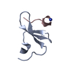
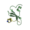
 Homo sapiens (human)
 X-RAY DIFFRACTION /
  Authors
 Citation
 Structure visualization
 Downloads & links
 Other downloads


 PDBj
PDBj


 Assembly
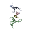

 Mass: 18.015 Da / Num. of mol.: 10 / Source method: isolated from a natural source / Formula: H2O
 Sample preparation
 Processing