 Movie
Movie Controller

[English] 日本語
 Yorodumi
- PDB-4rer: Crystal structure of the phosphorylated human alpha1 beta2 gamma1... -
+ Open data

- Basic information
| Entry | Database: PDB / ID: 4rer | |||||||||
|---|---|---|---|---|---|---|---|---|---|---|
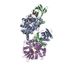
| Title | Crystal structure of the phosphorylated human alpha1 beta2 gamma1 holo-AMPK complex bound to AMP and cyclodextrin | |||||||||
 Components Components |
| |||||||||
 Keywords Keywords | TRANSFERASE / human alpha1 beta2 gamma1 holo-AMPK complex / serine/threonine protein kinase / axin / CaMKKbeta / LKB1 / glycogen / Phosphorylation | |||||||||
| Function / homology |  Function and homology information Function and homology informationnegative regulation of glucosylceramide biosynthetic process / positive regulation of mitochondrial transcription / [hydroxymethylglutaryl-CoA reductase (NADPH)] kinase / [hydroxymethylglutaryl-CoA reductase (NADPH)] kinase activity / regulation of stress granule assembly / AMPK inhibits chREBP transcriptional activation activity / negative regulation of tubulin deacetylation / histone H2BS36 kinase activity / cold acclimation / AMP-activated protein kinase activity ...negative regulation of glucosylceramide biosynthetic process / positive regulation of mitochondrial transcription / [hydroxymethylglutaryl-CoA reductase (NADPH)] kinase / [hydroxymethylglutaryl-CoA reductase (NADPH)] kinase activity / regulation of stress granule assembly / AMPK inhibits chREBP transcriptional activation activity / negative regulation of tubulin deacetylation / histone H2BS36 kinase activity / cold acclimation / AMP-activated protein kinase activity / lipid droplet disassembly / Lipophagy / regulation of carbon utilization / positive regulation of skeletal muscle tissue development / CAMKK-AMPK signaling cascade / cAMP-dependent protein kinase regulator activity / import into nucleus / regulation of vesicle-mediated transport / negative regulation of hepatocyte apoptotic process / Carnitine shuttle / Energy dependent regulation of mTOR by LKB1-AMPK / positive regulation of T cell mediated immune response to tumor cell / tau-protein kinase / nucleotide-activated protein kinase complex / protein kinase regulator activity / regulation of vascular permeability / negative regulation of TOR signaling / Activation of PPARGC1A (PGC-1alpha) by phosphorylation / protein localization to membrane / regulation of glycolytic process / cAMP-dependent protein kinase activity / : / protein localization to lipid droplet / response to caffeine / tau-protein kinase activity / cholesterol biosynthetic process / lipid biosynthetic process / cellular response to stress / AMP binding / Macroautophagy / fatty acid oxidation / motor behavior / negative regulation of ferroptosis / cellular response to ethanol / fatty acid homeostasis / negative regulation of lipid catabolic process / response to UV / cellular response to glucose starvation / positive regulation of protein localization / Activation of AMPK downstream of NMDARs / energy homeostasis / negative regulation of TORC1 signaling / negative regulation of insulin receptor signaling pathway / positive regulation of adipose tissue development / positive regulation of autophagy / cellular response to nutrient levels / positive regulation of gluconeogenesis / cellular response to calcium ion / positive regulation of glycolytic process / cellular response to starvation / response to activity / regulation of microtubule cytoskeleton organization / response to gamma radiation / protein localization to plasma membrane / TP53 Regulates Metabolic Genes / Translocation of SLC2A4 (GLUT4) to the plasma membrane / cellular response to glucose stimulus / neuron cellular homeostasis / ADP binding / regulation of circadian rhythm / positive regulation of cholesterol biosynthetic process / response to estrogen / tau protein binding / cellular response to xenobiotic stimulus / glucose metabolic process / autophagy / positive regulation of T cell activation / Wnt signaling pathway / fatty acid biosynthetic process / cellular response to hydrogen peroxide / rhythmic process / glucose homeostasis / positive regulation of cold-induced thermogenesis / cellular response to prostaglandin E stimulus / cellular response to oxidative stress / spermatogenesis / cellular response to hypoxia / Regulation of TP53 Activity through Phosphorylation / protein phosphorylation / response to hypoxia / protein kinase activity / non-specific serine/threonine protein kinase / regulation of cell cycle / apical plasma membrane / nuclear speck / cilium / ciliary basal body / negative regulation of gene expression / protein serine kinase activity / axon Similarity search - Function | |||||||||
| Biological species |  Homo sapiens (human) Homo sapiens (human) | |||||||||
| Method |  X-RAY DIFFRACTION / SYNCHROTRON / MOLECULAR REPLACEMENT / Resolution: 4.047 Å X-RAY DIFFRACTION / SYNCHROTRON / MOLECULAR REPLACEMENT / Resolution: 4.047 Å | |||||||||
 Authors Authors | Zhou, X.E. / Ke, J. / Li, X. / Wang, L. / Gu, X. / de Waal, P.W. / Tan, M.H.E. / Wang, D. / Wu, D. / Xu, H.E. / Melcher, K. | |||||||||
 Citation Citation | Journal: Cell Res. / Year: 2015 Title: Structural basis of AMPK regulation by adenine nucleotides and glycogen. Authors: Li, X. / Wang, L. / Zhou, X.E. / Ke, J. / de Waal, P.W. / Gu, X. / Tan, M.H. / Wang, D. / Wu, D. / Xu, H.E. / Melcher, K. | |||||||||
| History |
|
- Structure visualization
| Structure viewer | Molecule: MolmilJmol/JSmol |
|---|
- Downloads & links
-Download
| PDBx/mmCIF format | 4rer.cif.gz | 416.3 KB | Display | PDBx/mmCIF format |
|---|---|---|---|---|
| PDB format | pdb4rer.ent.gz | 340.6 KB | Display | PDB format |
| PDBx/mmJSON format | 4rer.json.gz | Tree view | PDBx/mmJSON format | |
| Others |  Other downloads Other downloads |
-Validation report
| Arichive directory | https://data.pdbj.org/pub/pdb/validation_reports/re/4rerftp://data.pdbj.org/pub/pdb/validation_reports/re/4rer | HTTPS FTP |
|---|
-Related structure data
| Related structure data |  4redC 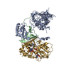 4rewC  2y94 C: citing same article ( S: Starting model for refinement |
|---|---|
| Similar structure data |


-Links
 PDBj
PDBj


- Assembly
| Deposited unit | 
| ||||||||
|---|---|---|---|---|---|---|---|---|---|
| 1 |
| ||||||||
| Unit cell |
| ||||||||
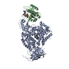
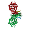
| Details | The heterotrimeric serine/threonine protein kinase |
-Components
-5'-AMP-activated protein kinase subunit ... , 2 types, 2 molecules BG
| #2: Protein | Mass: 22449.506 Da / Num. of mol.: 1 / Fragment: Human AMPK beta2 subunit [A76-I272] Source method: isolated from a genetically manipulated source Source: (gene. exp.) Homo sapiens (human) / Gene: Human holo-AMPK beta2 subunit, PRKAB2 / Plasmid: PET28 / Production host:  |
|---|---|
| #3: Protein | Mass: 34645.109 Da / Num. of mol.: 1 / Fragment: Human AMPK gamma1 subunit [S24-G327] Source method: isolated from a genetically manipulated source Source: (gene. exp.) Homo sapiens (human) / Gene: Human holo-AMPK gamma1 subunit, PRKAG1 / Plasmid: PET28 / Production host: |
-Protein / Sugars , 2 types, 2 molecules A
| #1: Protein | Mass: 61566.129 Da / Num. of mol.: 1 / Fragment: Human AMPK alpha1 subunit [G11-Q550] / Mutation: E471G, E474A, K476A Source method: isolated from a genetically manipulated source Source: (gene. exp.) Homo sapiens (human) / Gene: AMPK1, Human holo-AMPK alpha1 subunit, PRKAA1 / Plasmid: PET28 / Production host: References: UniProt: Q13131, non-specific serine/threonine protein kinase, EC: 2.7.11.27, [hydroxymethylglutaryl-CoA reductase (NADPH)] kinase, tau-protein kinase |
|---|---|
| #4: Polysaccharide | Cycloheptakis-(1-4)-(alpha-D-glucopyranose) / beta-cyclodextrin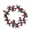  Source method: isolated from a genetically manipulated source Details: cyclic oligosaccharide / References: beta-cyclodextrin |
-Non-polymers , 3 types, 5 molecules 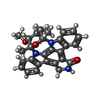


| #5: Chemical | ChemComp-STU /  Mass: 466.531 Da / Num. of mol.: 1 / Source method: obtained synthetically / Formula: C28H26N4O3 / Comment: anticancer, antifungal, antibiotic, alkaloid*YM Mass: 466.531 Da / Num. of mol.: 1 / Source method: obtained synthetically / Formula: C28H26N4O3 / Comment: anticancer, antifungal, antibiotic, alkaloid*YM |
|---|---|
| #6: Chemical | ChemComp-EPE /  Mass: 238.305 Da / Num. of mol.: 1 / Source method: obtained synthetically / Formula: C8H18N2O4S / Comment: pH buffer*YM Mass: 238.305 Da / Num. of mol.: 1 / Source method: obtained synthetically / Formula: C8H18N2O4S / Comment: pH buffer*YM |
| #7: Chemical |  Mass: 347.221 Da / Num. of mol.: 3 / Source method: obtained synthetically / Formula: C10H14N5O7P / Comment: AMP*YM Mass: 347.221 Da / Num. of mol.: 3 / Source method: obtained synthetically / Formula: C10H14N5O7P / Comment: AMP*YM |
-Details
| Has protein modification | Y |
|---|
-Experimental details
-Experiment
| Experiment | Method: X-RAY DIFFRACTION / Number of used crystals: 1 |
|---|
- Sample preparation
| Crystal | Density Matthews: 4.18 Å3/Da / Density % sol: 70.55 % |
|---|---|
| Crystal grow | Temperature: 293 K / Method: vapor diffusion, sitting drop / pH: 7.8 Details: 12% PEG 4000,0.1 M HEPES, pH7.8, 10% 2-propanol (v/v) and 0.19 mM 7-cyclohexyl-1-heptyl-D-maltoside, VAPOR DIFFUSION, SITTING DROP, temperature 293K |
-Data collection
| Diffraction | Mean temperature: 100 K |
|---|---|
| Diffraction source | Source: SYNCHROTRON / Site: APS  / Beamline: 21-ID-F / Wavelength: 0.97872 Å / Beamline: 21-ID-F / Wavelength: 0.97872 Å |
| Detector | Type: MARMOSAIC 225 mm CCD / Detector: CCD / Date: Jul 19, 2012 |
| Radiation | Monochromator: Ni FILTER / Protocol: SINGLE WAVELENGTH / Monochromatic (M) / Laue (L): M / Scattering type: x-ray |
| Radiation wavelength | Wavelength: 0.97872 Å / Relative weight: 1 |
| Reflection | Resolution: 4.05→40 Å / Num. all: 16738 / Num. obs: 16621 / % possible obs: 99.3 % / Redundancy: 13.1 % / Rmerge(I) obs: 0.186 / Net I/σ(I): 7.6 |
| Reflection shell | Resolution: 4.05→4.19 Å / % possible all: 93 |
- Processing
| Software |
| |||||||||||||||||||||||||||||||||||||||||||||||||||||||||||||||||||||||||||||||||||||||||||||||||||||||||||||||||||||||||||||
|---|---|---|---|---|---|---|---|---|---|---|---|---|---|---|---|---|---|---|---|---|---|---|---|---|---|---|---|---|---|---|---|---|---|---|---|---|---|---|---|---|---|---|---|---|---|---|---|---|---|---|---|---|---|---|---|---|---|---|---|---|---|---|---|---|---|---|---|---|---|---|---|---|---|---|---|---|---|---|---|---|---|---|---|---|---|---|---|---|---|---|---|---|---|---|---|---|---|---|---|---|---|---|---|---|---|---|---|---|---|---|---|---|---|---|---|---|---|---|---|---|---|---|---|---|---|---|
| Refinement | Method to determine structure: MOLECULAR REPLACEMENT Starting model: PDB ENTRY 2Y94 2y94 Resolution: 4.047→39.657 Å / SU ML: 0.55 / σ(F): 1.35 / Phase error: 28.23 / Stereochemistry target values: MAXIMUM-LIKELIHOOD
| |||||||||||||||||||||||||||||||||||||||||||||||||||||||||||||||||||||||||||||||||||||||||||||||||||||||||||||||||||||||||||||
| Solvent computation | Shrinkage radii: 0.9 Å / VDW probe radii: 1.11 Å / Solvent model: FLAT BULK SOLVENT MODEL | |||||||||||||||||||||||||||||||||||||||||||||||||||||||||||||||||||||||||||||||||||||||||||||||||||||||||||||||||||||||||||||
| Refinement step | Cycle: LAST / Resolution: 4.047→39.657 Å
| |||||||||||||||||||||||||||||||||||||||||||||||||||||||||||||||||||||||||||||||||||||||||||||||||||||||||||||||||||||||||||||
| Refine LS restraints |
| |||||||||||||||||||||||||||||||||||||||||||||||||||||||||||||||||||||||||||||||||||||||||||||||||||||||||||||||||||||||||||||
| LS refinement shell |
| |||||||||||||||||||||||||||||||||||||||||||||||||||||||||||||||||||||||||||||||||||||||||||||||||||||||||||||||||||||||||||||
| Refinement TLS params. | Method: refined / Refine-ID: X-RAY DIFFRACTION
| |||||||||||||||||||||||||||||||||||||||||||||||||||||||||||||||||||||||||||||||||||||||||||||||||||||||||||||||||||||||||||||
| Refinement TLS group |
|
