 Movie
Movie Controller

[English] 日本語
 Yorodumi
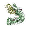
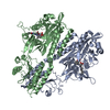
- PDB-4r6l: Crystal structure of bacteriophytochrome RpBphP2 from photosynthe... -
+ Open data

- Basic information
| Entry | Database: PDB / ID: 4r6l | ||||||
|---|---|---|---|---|---|---|---|
| Title | Crystal structure of bacteriophytochrome RpBphP2 from photosynthetic bacterium R. palustris | ||||||
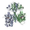
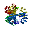
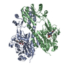
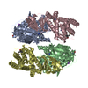
 Components Components | Bacteriophytochrome (Light-regulated signal transduction histidine kinase), PhyB1 | ||||||
 Keywords Keywords | SIGNALING PROTEIN / PAS fold / photoreceptor / response regulator RPA3017 / TRANSFERASE | ||||||
| Function / homology |  Function and homology information Function and homology informationosmosensory signaling via phosphorelay pathway / detection of visible light / phosphorelay response regulator activity / phosphorelay sensor kinase activity / histidine kinase / photoreceptor activity / protein kinase activator activity / regulation of DNA-templated transcription / ATP binding / metal ion binding Similarity search - Function | ||||||
| Biological species |  Rhodopseudomonas palustris CGA009 (phototrophic) Rhodopseudomonas palustris CGA009 (phototrophic) | ||||||
| Method |  X-RAY DIFFRACTION / SYNCHROTRON / MOLECULAR REPLACEMENT / Resolution: 3.395 Å X-RAY DIFFRACTION / SYNCHROTRON / MOLECULAR REPLACEMENT / Resolution: 3.395 Å | ||||||
 Authors Authors | Yang, X. / Stojkovic, E. / Ozarowski, W. / Kuk, J. / Davydova, E. / Moffat, K. | ||||||
 Citation Citation | Journal: Structure / Year: 2015 Title: Light Signaling Mechanism of Two Tandem Bacteriophytochromes. Authors: Yang, X. / Stojkovic, E.A. / Ozarowski, W.B. / Kuk, J. / Davydova, E. / Moffat, K. | ||||||
| History |
|
- Structure visualization
| Structure viewer | Molecule: MolmilJmol/JSmol |
|---|
- Downloads & links
-Download
| PDBx/mmCIF format | 4r6l.cif.gz | 378.6 KB | Display | PDBx/mmCIF format |
|---|---|---|---|---|
| PDB format | pdb4r6l.ent.gz | 316.8 KB | Display | PDB format |
| PDBx/mmJSON format | 4r6l.json.gz | Tree view | PDBx/mmJSON format | |
| Others |  Other downloads Other downloads |
-Validation report
| Arichive directory | https://data.pdbj.org/pub/pdb/validation_reports/r6/4r6lftp://data.pdbj.org/pub/pdb/validation_reports/r6/4r6l | HTTPS FTP |
|---|
-Related structure data
| Related structure data |  4r70C  4s21C 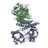 2oolS S: Starting model for refinement C: citing same article ( |
|---|---|
| Similar structure data |
-Links
 PDBj
PDBj


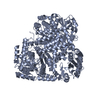
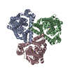
- Assembly
| Deposited unit | 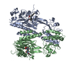
| ||||||||
|---|---|---|---|---|---|---|---|---|---|
| 1 |
| ||||||||
| Unit cell |
|
-Components
| #1: Protein | Mass: 56814.555 Da / Num. of mol.: 2 / Fragment: photosensory core module, UNP residues 1-506 Source method: isolated from a genetically manipulated source Source: (gene. exp.) Rhodopseudomonas palustris CGA009 (phototrophic)Strain: CGA009 / Gene: phyB1, RPA3015 / Plasmid: pET24 / Production host: #2: Chemical |   Mass: 582.646 Da / Num. of mol.: 2 / Source method: obtained synthetically / Formula: C33H34N4O6 Mass: 582.646 Da / Num. of mol.: 2 / Source method: obtained synthetically / Formula: C33H34N4O6Has protein modification | Y | |
|---|
-Experimental details
-Experiment
| Experiment | Method: X-RAY DIFFRACTION / Number of used crystals: 1 |
|---|
- Sample preparation
| Crystal | Density Matthews: 3.81 Å3/Da / Density % sol: 67.7 % |
|---|---|
| Crystal grow | Temperature: 293 K / Method: vapor diffusion, hanging drop / pH: 7 Details: 10mg/ml protein, 12% polyacrylic acid, 0.1M MgCl2, 0.1M Hepes (pH7.0), VAPOR DIFFUSION, HANGING DROP, temperature 293K |
-Data collection
| Diffraction | Mean temperature: 100 K |
|---|---|
| Diffraction source | Source: SYNCHROTRON / Site: APS  / Beamline: 21-ID-G / Wavelength: 0.97857 Å / Beamline: 21-ID-G / Wavelength: 0.97857 Å |
| Detector | Type: RAYONIX MX-300 / Detector: CCD / Date: Feb 8, 2009 |
| Radiation | Monochromator: diamond laue monochromators / Protocol: SINGLE WAVELENGTH / Monochromatic (M) / Laue (L): M / Scattering type: x-ray |
| Radiation wavelength | Wavelength: 0.97857 Å / Relative weight: 1 |
| Reflection | Resolution: 3.306→50 Å / Num. obs: 22049 / % possible obs: 62 % / Observed criterion σ(F): 0 / Observed criterion σ(I): 0 / Redundancy: 4.5 % / Rmerge(I) obs: 0.057 / Net I/σ(I): 15 |
- Processing
| Software |
| |||||||||||||||||||||||||||||||||||||||||||||||||||||||||||||||||||||||||||
|---|---|---|---|---|---|---|---|---|---|---|---|---|---|---|---|---|---|---|---|---|---|---|---|---|---|---|---|---|---|---|---|---|---|---|---|---|---|---|---|---|---|---|---|---|---|---|---|---|---|---|---|---|---|---|---|---|---|---|---|---|---|---|---|---|---|---|---|---|---|---|---|---|---|---|---|---|
| Refinement | Method to determine structure: MOLECULAR REPLACEMENT Starting model: PDB ENTRY 2OOL Resolution: 3.395→49.021 Å / SU ML: 0.66 / σ(F): 1.34 / Phase error: 41.64 / Stereochemistry target values: ML Details: Author states that issues with the main chain geometry and the high B-factors are largely due to the poor resolution.
| |||||||||||||||||||||||||||||||||||||||||||||||||||||||||||||||||||||||||||
| Solvent computation | Shrinkage radii: 0.9 Å / VDW probe radii: 1.11 Å / Solvent model: FLAT BULK SOLVENT MODEL | |||||||||||||||||||||||||||||||||||||||||||||||||||||||||||||||||||||||||||
| Refinement step | Cycle: LAST / Resolution: 3.395→49.021 Å
| |||||||||||||||||||||||||||||||||||||||||||||||||||||||||||||||||||||||||||
| Refine LS restraints |
| |||||||||||||||||||||||||||||||||||||||||||||||||||||||||||||||||||||||||||
| LS refinement shell |
| |||||||||||||||||||||||||||||||||||||||||||||||||||||||||||||||||||||||||||
| Refinement TLS params. | Method: refined / Refine-ID: X-RAY DIFFRACTION
| |||||||||||||||||||||||||||||||||||||||||||||||||||||||||||||||||||||||||||
| Refinement TLS group |
|
