purine ribonucleoside triphosphate binding / CD8-positive, alpha-beta T cell differentiation / thymic T cell selection / CD8-positive, alpha-beta T cell homeostasis / Antigen processing: Ub, ATP-independent proteasomal degradation / T-helper 1 cell differentiation / negative regulation of regulatory T cell differentiation / cellular response to type I interferon / Regulation of ornithine decarboxylase (ODC) / proteasome core complex ...purine ribonucleoside triphosphate binding / CD8-positive, alpha-beta T cell differentiation / thymic T cell selection / CD8-positive, alpha-beta T cell homeostasis / Antigen processing: Ub, ATP-independent proteasomal degradation / T-helper 1 cell differentiation / negative regulation of regulatory T cell differentiation / cellular response to type I interferon / Regulation of ornithine decarboxylase (ODC) / proteasome core complex / Proteasome assembly / T-helper 17 cell differentiation / Cross-presentation of soluble exogenous antigens (endosomes) / Somitogenesis / flagellated sperm motility / myofibril / AMPK-induced ERAD and lysosome mediated degradation of PD-L1(CD274) / GSK3B-mediated proteasomal degradation of PD-L1(CD274) / SPOP-mediated proteasomal degradation of PD-L1(CD274) / proteasomal ubiquitin-independent protein catabolic process / Ribosome Quality Control (RQC) complex extracts and degrades nascent peptide / proteasome storage granule / proteasome endopeptidase complex / NF-kappaB binding / proteasome core complex, beta-subunit complex / threonine-type endopeptidase activity / proteasome core complex, alpha-subunit complex / proteasome assembly / immune system process / regulation of G1/S transition of mitotic cell cycle / ciliary tip / response to type II interferon / positive regulation of interleukin-2 production / regulation of proteasomal protein catabolic process / : / proteasome complex / sarcomere / proteasomal protein catabolic process / Regulation of activated PAK-2p34 by proteasome mediated degradation / sperm end piece / Autodegradation of Cdh1 by Cdh1:APC/C / APC/C:Cdc20 mediated degradation of Securin / negative regulation of inflammatory response to antigenic stimulus / Asymmetric localization of PCP proteins / Ubiquitin-dependent degradation of Cyclin D / lipopolysaccharide binding / SCF-beta-TrCP mediated degradation of Emi1 / NIK-->noncanonical NF-kB signaling / AUF1 (hnRNP D0) binds and destabilizes mRNA / TNFR2 non-canonical NF-kB pathway / Assembly of the pre-replicative complex / Vpu mediated degradation of CD4 / P-body / Cdc20:Phospho-APC/C mediated degradation of Cyclin A / Dectin-1 mediated noncanonical NF-kB signaling / Degradation of DVL / Degradation of AXIN / Degradation of CRY and PER proteins / meiotic cell cycle / Hh mutants are degraded by ERAD / Activation of NF-kappaB in B cells / G2/M Checkpoints / Degradation of GLI1 by the proteasome / Hedgehog ligand biogenesis / Regulation of RUNX3 expression and activity / Autodegradation of the E3 ubiquitin ligase COP1 / Defective CFTR causes cystic fibrosis / GSK3B and BTRC:CUL1-mediated-degradation of NFE2L2 / Negative regulation of NOTCH4 signaling / APC/C:Cdh1 mediated degradation of Cdc20 and other APC/C:Cdh1 targeted proteins in late mitosis/early G1 / Hedgehog 'on' state / FBXL7 down-regulates AURKA during mitotic entry and in early mitosis / Vif-mediated degradation of APOBEC3G / Degradation of GLI2 by the proteasome / GLI3 is processed to GLI3R by the proteasome / Ubiquitin-Mediated Degradation of Phosphorylated Cdc25A / MAPK6/MAPK4 signaling / Degradation of CDH1 / Degradation of beta-catenin by the destruction complex / Oxygen-dependent proline hydroxylation of Hypoxia-inducible Factor Alpha / CDK-mediated phosphorylation and removal of Cdc6 / ABC-family protein mediated transport / CLEC7A (Dectin-1) signaling / response to virus / SCF(Skp2)-mediated degradation of p27/p21 / FCERI mediated NF-kB activation / nuclear matrix / Regulation of expression of SLITs and ROBOs / Regulation of PTEN stability and activity / Interleukin-1 signaling / positive regulation of type II interferon production / Orc1 removal from chromatin / Regulation of RUNX2 expression and activity / Regulation of RAS by GAPs / The role of GTSE1 in G2/M progression after G2 checkpoint / positive regulation of tumor necrosis factor production / Separation of Sister Chromatids / UCH proteinases / KEAP1-NFE2L2 pathway / peptidase activity Similarity search - Function
 Movie
Movie Controller


 Open data
 Basic information
 Components
 Keywords
 Function and homology information
 Homo sapiens (human)
 X-RAY DIFFRACTION /
  Authors
 Citation
 Structure visualization
 Downloads & links
 Other downloads


 PDBj
PDBj


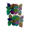
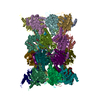
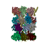
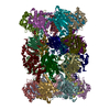
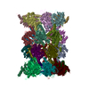
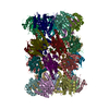
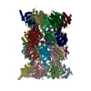
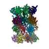
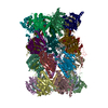
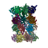
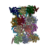
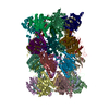
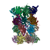
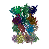
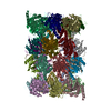
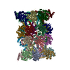
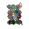
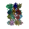
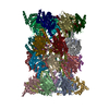
 Assembly