 Movie
Movie Controller


+ Open data

- Basic information
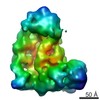
| Entry | Database: PDB / ID: 4r2y | ||||||
|---|---|---|---|---|---|---|---|
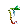
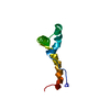
| Title | Crystal structure of APC11 RING domain | ||||||
 Components Components | Anaphase-promoting complex subunit 11 | ||||||
 Keywords Keywords | LIGASE / RING domain / E3 Ubiquitin Ligase | ||||||
| Function / homology |  Function and homology information Function and homology informationConversion from APC/C:Cdc20 to APC/C:Cdh1 in late anaphase / Inactivation of APC/C via direct inhibition of the APC/C complex / APC/C:Cdc20 mediated degradation of mitotic proteins / anaphase-promoting complex / anaphase-promoting complex-dependent catabolic process / Aberrant regulation of mitotic exit in cancer due to RB1 defects / regulation of meiotic cell cycle / protein branched polyubiquitination / Phosphorylation of the APC/C / positive regulation of mitotic metaphase/anaphase transition ...Conversion from APC/C:Cdc20 to APC/C:Cdh1 in late anaphase / Inactivation of APC/C via direct inhibition of the APC/C complex / APC/C:Cdc20 mediated degradation of mitotic proteins / anaphase-promoting complex / anaphase-promoting complex-dependent catabolic process / Aberrant regulation of mitotic exit in cancer due to RB1 defects / regulation of meiotic cell cycle / protein branched polyubiquitination / Phosphorylation of the APC/C / positive regulation of mitotic metaphase/anaphase transition / protein K11-linked ubiquitination / ubiquitin-ubiquitin ligase activity / Regulation of APC/C activators between G1/S and early anaphase / Transcriptional Regulation by VENTX / cullin family protein binding / protein K48-linked ubiquitination / regulation of mitotic cell cycle / APC/C:Cdc20 mediated degradation of Cyclin B / APC-Cdc20 mediated degradation of Nek2A / Autodegradation of Cdh1 by Cdh1:APC/C / APC/C:Cdc20 mediated degradation of Securin / Assembly of the pre-replicative complex / Cdc20:Phospho-APC/C mediated degradation of Cyclin A / APC/C:Cdh1 mediated degradation of Cdc20 and other APC/C:Cdh1 targeted proteins in late mitosis/early G1 / G protein-coupled receptor binding / CDK-mediated phosphorylation and removal of Cdc6 / ubiquitin-protein transferase activity / Separation of Sister Chromatids / ubiquitin protein ligase activity / mitotic cell cycle / Antigen processing: Ubiquitination & Proteasome degradation / Senescence-Associated Secretory Phenotype (SASP) / ubiquitin-dependent protein catabolic process / protein ubiquitination / cell division / nucleolus / nucleoplasm / zinc ion binding / nucleus / cytosol Similarity search - Function | ||||||
| Biological species |  Homo sapiens (human) Homo sapiens (human) | ||||||
| Method |  X-RAY DIFFRACTION / SYNCHROTRON / SAD / Resolution: 1.755 Å X-RAY DIFFRACTION / SYNCHROTRON / SAD / Resolution: 1.755 Å | ||||||
 Authors Authors | Brown, N.G. / Watson, E.R. / Weissmann, F. / Jarvis, M.A. / Vanderlinden, R. / Grace, C.R.R. / Frye, J.J. / Dube, P. / Qiao, R. / Petzold, G. ...Brown, N.G. / Watson, E.R. / Weissmann, F. / Jarvis, M.A. / Vanderlinden, R. / Grace, C.R.R. / Frye, J.J. / Dube, P. / Qiao, R. / Petzold, G. / Cho, S.E. / Alsharif, O. / Bao, J. / Zheng, J. / Nourse, A. / Kurinov, I. / Peters, J.M. / Stark, H. / Schulman, B.A. | ||||||
 Citation Citation | Journal: Mol Cell / Year: 2014 Title: Mechanism of polyubiquitination by human anaphase-promoting complex: RING repurposing for ubiquitin chain assembly. Authors: Nicholas G Brown / Edmond R Watson / Florian Weissmann / Marc A Jarvis / Ryan VanderLinden / Christy R R Grace / Jeremiah J Frye / Renping Qiao / Prakash Dube / Georg Petzold / Shein Ei Cho ...Authors: Nicholas G Brown / Edmond R Watson / Florian Weissmann / Marc A Jarvis / Ryan VanderLinden / Christy R R Grace / Jeremiah J Frye / Renping Qiao / Prakash Dube / Georg Petzold / Shein Ei Cho / Omar Alsharif / Ju Bao / Iain F Davidson / Jie J Zheng / Amanda Nourse / Igor Kurinov / Jan-Michael Peters / Holger Stark / Brenda A Schulman /    Abstract: Polyubiquitination by E2 and E3 enzymes is a predominant mechanism regulating protein function. Some RING E3s, including anaphase-promoting complex/cyclosome (APC), catalyze polyubiquitination by ...Polyubiquitination by E2 and E3 enzymes is a predominant mechanism regulating protein function. Some RING E3s, including anaphase-promoting complex/cyclosome (APC), catalyze polyubiquitination by sequential reactions with two different E2s. An initiating E2 ligates ubiquitin to an E3-bound substrate. Another E2 grows a polyubiquitin chain on the ubiquitin-primed substrate through poorly defined mechanisms. Here we show that human APC's RING domain is repurposed for dual functions in polyubiquitination. The canonical RING surface activates an initiating E2-ubiquitin intermediate for substrate modification. However, APC engages and activates its specialized ubiquitin chain-elongating E2 UBE2S in ways that differ from current paradigms. During chain assembly, a distinct APC11 RING surface helps deliver a substrate-linked ubiquitin to accept another ubiquitin from UBE2S. Our data define mechanisms of APC/UBE2S-mediated polyubiquitination, reveal diverse functions of RING E3s and E2s, and provide a framework for understanding distinctive RING E3 features specifying ubiquitin chain elongation. | ||||||
| History |
|
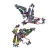
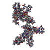
- Structure visualization
| Structure viewer | Molecule: MolmilJmol/JSmol |
|---|
- Downloads & links
-Download
| PDBx/mmCIF format | 4r2y.cif.gz | 122.3 KB | Display | PDBx/mmCIF format |
|---|---|---|---|---|
| PDB format | pdb4r2y.ent.gz | 96.2 KB | Display | PDB format |
| PDBx/mmJSON format | 4r2y.json.gz | Tree view | PDBx/mmJSON format | |
| Others |  Other downloads Other downloads |
-Validation report
| Arichive directory | https://data.pdbj.org/pub/pdb/validation_reports/r2/4r2yftp://data.pdbj.org/pub/pdb/validation_reports/r2/4r2y | HTTPS FTP |
|---|
-Related structure data


-Links
 PDBj
PDBj


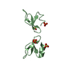
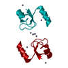
- Assembly
| Deposited unit | 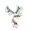
| ||||||||
|---|---|---|---|---|---|---|---|---|---|
| 1 | 
| ||||||||
| 2 | 
| ||||||||
| 3 | 
| ||||||||
| 4 | 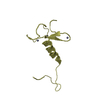
| ||||||||
| 5 | 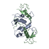
| ||||||||
| 6 | 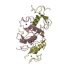
| ||||||||
| Unit cell |
| ||||||||
| Details | The structure is of a domain-swapped dimer. The domain swap occurs at VAL 69. To generate the biological unit, it is necessary to pair residues 21-68 from Chain A with 69-84 of Chain B, residues 21-68 from Chain B with 69-84 of Chain A, residues 20-68 of Chain C with 69-84 of Chain D, and 20-68 of Chain D with 69-84 of Chain C. |
-Components
| #1: Protein | Mass: 7906.229 Da / Num. of mol.: 4 / Fragment: RING domain (UNP Residues 17-84) Source method: isolated from a genetically manipulated source Source: (gene. exp.) Homo sapiens (human) / Gene: ANAPC11, HSPC214 / Production host:  #2: Chemical | ChemComp-ZN /   Mass: 65.409 Da / Num. of mol.: 12 / Source method: obtained synthetically / Formula: Zn Mass: 65.409 Da / Num. of mol.: 12 / Source method: obtained synthetically / Formula: Zn#3: Water | ChemComp-HOH / |  Mass: 18.015 Da / Num. of mol.: 208 / Source method: isolated from a natural source / Formula: H2O Mass: 18.015 Da / Num. of mol.: 208 / Source method: isolated from a natural source / Formula: H2O |
|---|
-Experimental details
-Experiment
| Experiment | Method: X-RAY DIFFRACTION / Number of used crystals: 1 |
|---|
- Sample preparation
| Crystal | Density Matthews: 2.04 Å3/Da / Density % sol: 39.69 % |
|---|---|
| Crystal grow | Temperature: 298 K / Method: vapor diffusion, hanging drop / pH: 6.5 Details: 16% PEG3350, 0.2 M NaNO3, 0.1 M Bis-Tris, pH 6.5, VAPOR DIFFUSION, HANGING DROP, temperature 298K |
-Data collection
| Diffraction | Mean temperature: 100 K |
|---|---|
| Diffraction source | Source: SYNCHROTRON / Site: APS / Beamline: 24-ID-C / Wavelength: 1.2827 Å |
| Detector | Type: DECTRIS PILATUS 6M-F / Detector: PIXEL / Date: Jul 1, 2014 |
| Radiation | Monochromator: CRYO-COOLED DOUBLE CRYSTAL / Protocol: SINGLE WAVELENGTH / Monochromatic (M) / Laue (L): M / Scattering type: x-ray |
| Radiation wavelength | Wavelength: 1.2827 Å / Relative weight: 1 |
| Reflection | Resolution: 1.755→61.681 Å / Num. obs: 24444 / % possible obs: 94.59 % / Observed criterion σ(I): 2 |
| Reflection shell | Resolution: 1.755→1.85 Å / % possible all: 80.2 |
- Processing
| Software |
| |||||||||||||||||||||||||||||||||||||||||||||||||||||||||||||||||||||||||||||||||||||||||||||||||||||||||
|---|---|---|---|---|---|---|---|---|---|---|---|---|---|---|---|---|---|---|---|---|---|---|---|---|---|---|---|---|---|---|---|---|---|---|---|---|---|---|---|---|---|---|---|---|---|---|---|---|---|---|---|---|---|---|---|---|---|---|---|---|---|---|---|---|---|---|---|---|---|---|---|---|---|---|---|---|---|---|---|---|---|---|---|---|---|---|---|---|---|---|---|---|---|---|---|---|---|---|---|---|---|---|---|---|---|---|
| Refinement | Method to determine structure: SAD / Resolution: 1.755→61.681 Å / SU ML: 0.46 / Phase error: 21.9 / Stereochemistry target values: ML
| |||||||||||||||||||||||||||||||||||||||||||||||||||||||||||||||||||||||||||||||||||||||||||||||||||||||||
| Solvent computation | Shrinkage radii: 0.61 Å / VDW probe radii: 0.9 Å / Solvent model: FLAT BULK SOLVENT MODEL / Bsol: 40.012 Å2 / ksol: 0.401 e/Å3 | |||||||||||||||||||||||||||||||||||||||||||||||||||||||||||||||||||||||||||||||||||||||||||||||||||||||||
| Displacement parameters |
| |||||||||||||||||||||||||||||||||||||||||||||||||||||||||||||||||||||||||||||||||||||||||||||||||||||||||
| Refinement step | Cycle: LAST / Resolution: 1.755→61.681 Å
| |||||||||||||||||||||||||||||||||||||||||||||||||||||||||||||||||||||||||||||||||||||||||||||||||||||||||
| Refine LS restraints |
| |||||||||||||||||||||||||||||||||||||||||||||||||||||||||||||||||||||||||||||||||||||||||||||||||||||||||
| LS refinement shell |
| |||||||||||||||||||||||||||||||||||||||||||||||||||||||||||||||||||||||||||||||||||||||||||||||||||||||||
| Refinement TLS params. | Method: refined / Origin x: 43.5023 Å / Origin y: 1.6788 Å / Origin z: 15.4601 Å
| |||||||||||||||||||||||||||||||||||||||||||||||||||||||||||||||||||||||||||||||||||||||||||||||||||||||||
| Refinement TLS group | Selection details: all |
