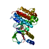
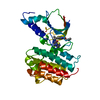
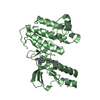
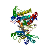
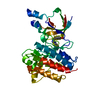
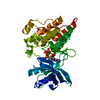
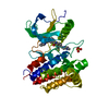
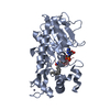
Entry Database : PDB / ID : 4r1vTitle Identification and optimization of pyridazinones as potent and selective c-Met kinase inhibitors Hepatocyte growth factor receptor Keywords / / / Function / homology Function Domain/homology Component
/ / / / / / / / / / / / / / / / / / / / / / / / / / / / / / / / / / / / / / / / / / / / / / / / / / / / / / / / / / / / / / / / / / / / / / / / / / / / / / / / / / / / / / / / / / / / / / / / / / / / / Biological species Homo sapiens (human)Method / / Resolution : 1.2 Å Authors Blaukat, A. / Bladt, F. / Friese-Hamim, M. / Knuehl, C. / Fittschen, C. / Graedler, U. / Meyring, M. / Dorsch, D. / Stieber, F. / Schadt, O. Journal : Bioorg.Med.Chem.Lett. / Year : 2015Title : Identification and optimization of pyridazinones as potent and selective c-Met kinase inhibitors.Authors : Dorsch, D. / Schadt, O. / Stieber, F. / Meyring, M. / Gradler, U. / Bladt, F. / Friese-Hamim, M. / Knuhl, C. / Pehl, U. / Blaukat, A. History Deposition Aug 7, 2014 Deposition site / Processing site Revision 1.0 Mar 18, 2015 Provider / Type Revision 1.1 Apr 22, 2015 Group Revision 1.2 Feb 28, 2024 Group / Database references / Derived calculationsCategory chem_comp_atom / chem_comp_bond ... chem_comp_atom / chem_comp_bond / database_2 / struct_site Item _database_2.pdbx_DOI / _database_2.pdbx_database_accession ... _database_2.pdbx_DOI / _database_2.pdbx_database_accession / _struct_site.pdbx_auth_asym_id / _struct_site.pdbx_auth_comp_id / _struct_site.pdbx_auth_seq_id
Show all Show less
 Movie
Movie Controller

 Yorodumi
 Open data
 Basic information
 Components
 Keywords
 Function and homology information
 Homo sapiens (human)
 X-RAY DIFFRACTION /
  Authors
 Citation
 Structure visualization
 Downloads & links
 Other downloads
 PDBj
PDBj


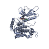
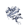
 Assembly
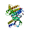

 Spodoptera frugiperda (fall armyworm)
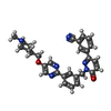
 Mass: 492.572 Da / Num. of mol.: 1 / Source method: obtained synthetically / Formula: C29H28N6O2
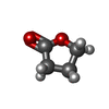
 Mass: 86.089 Da / Num. of mol.: 3 / Source method: obtained synthetically / Formula: C4H6O2
 Mass: 18.015 Da / Num. of mol.: 349 / Source method: isolated from a natural source / Formula: H2O
 Sample preparation
 / Beamline: X06DA / Wavelength: 0.9999
  Processing