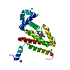
 Movie
Movie Controller


+ Open data

- Basic information
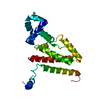
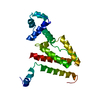
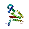
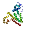
| Entry | Database: PDB / ID: 4qjn | ||||||
|---|---|---|---|---|---|---|---|
| Title | Crystal structure of apo nucleoid associated protein, SAV1473 | ||||||
 Components Components | DNA-binding protein HU | ||||||
 Keywords Keywords | DNA BINDING PROTEIN / DNA condensation / DNA binding | ||||||
| Function / homology |  Function and homology information Function and homology informationHU-DNA complex / DnaA-HU complex / chromosome condensation / DNA replication initiation / structural constituent of chromatin / gene expression / DNA binding / identical protein binding / cytosol Similarity search - Function | ||||||
| Biological species |   Staphylococcus aureus (bacteria) Staphylococcus aureus (bacteria) | ||||||
| Method |  X-RAY DIFFRACTION / SYNCHROTRON / MOLECULAR REPLACEMENT / Resolution: 2.613 Å X-RAY DIFFRACTION / SYNCHROTRON / MOLECULAR REPLACEMENT / Resolution: 2.613 Å | ||||||
 Authors Authors | Lee, B.-J. / Kim, D.-H. / Im, H. / Yoon, H.-J. | ||||||
 Citation Citation | Journal: Acta Crystallogr.,Sect.D / Year: 2014 Title: beta-Arm flexibility of HU from Staphylococcus aureus dictates the DNA-binding and recognition mechanism Authors: Kim, D.-H. / Im, H. / Jee, J.-G. / Jang, S.-B. / Yoon, H.-J. / Kwon, A.-R. / Kang, S.-M. / Lee, B.-J. | ||||||
| History |
|
- Structure visualization
| Structure viewer | Molecule: MolmilJmol/JSmol |
|---|
- Downloads & links
-Download
| PDBx/mmCIF format | 4qjn.cif.gz | 78.4 KB | Display | PDBx/mmCIF format |
|---|---|---|---|---|
| PDB format | pdb4qjn.ent.gz | 61 KB | Display | PDB format |
| PDBx/mmJSON format | 4qjn.json.gz | Tree view | PDBx/mmJSON format | |
| Others |  Other downloads Other downloads |
-Validation report
| Arichive directory | https://data.pdbj.org/pub/pdb/validation_reports/qj/4qjnftp://data.pdbj.org/pub/pdb/validation_reports/qj/4qjn | HTTPS FTP |
|---|
-Related structure data
| Related structure data |  4qjuC  1huuS C: citing same article ( S: Starting model for refinement |
|---|---|
| Similar structure data |


-Links
 PDBj
PDBj- Assembly
| Deposited unit | 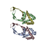
| ||||||||
|---|---|---|---|---|---|---|---|---|---|
| 1 | 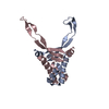
| ||||||||
| 2 | 
| ||||||||
| Unit cell |
|
-Components
| #1: Protein | Mass: 10713.167 Da / Num. of mol.: 4 Source method: isolated from a genetically manipulated source Source: (gene. exp.) Staphylococcus aureus (bacteria) / Strain: Mu50 / Gene: hup / Plasmid: pET21a / Production host: #2: Water | ChemComp-HOH / |  Mass: 18.015 Da / Num. of mol.: 59 / Source method: isolated from a natural source / Formula: H2O Mass: 18.015 Da / Num. of mol.: 59 / Source method: isolated from a natural source / Formula: H2O |
|---|
-Experimental details
-Experiment
| Experiment | Method: X-RAY DIFFRACTION / Number of used crystals: 1 |
|---|
- Sample preparation
| Crystal | Density Matthews: 2.14 Å3/Da / Density % sol: 42.63 % |
|---|---|
| Crystal grow | Temperature: 293 K / Method: vapor diffusion, hanging drop Details: PEG2000 monomethyl ether, VAPOR DIFFUSION, HANGING DROP, temperature 293K |
-Data collection
| Diffraction | Mean temperature: 100 K |
|---|---|
| Diffraction source | Source: SYNCHROTRON / Site: SPring-8  / Beamline: BL26B1 / Wavelength: 1 Å / Beamline: BL26B1 / Wavelength: 1 Å |
| Detector | Type: RIGAKU SATURN A200 / Detector: CCD / Date: May 18, 2011 |
| Radiation | Monochromator: Fixed exit Si double crystal monochromator / Protocol: SINGLE WAVELENGTH / Monochromatic (M) / Laue (L): M / Scattering type: x-ray |
| Radiation wavelength | Wavelength: 1 Å / Relative weight: 1 |
| Reflection | Resolution: 2.61→50 Å / Num. obs: 10877 / Redundancy: 6 % / Biso Wilson estimate: 41.6 Å2 / Rmerge(I) obs: 0.083 / Net I/σ(I): 33.7 |
| Reflection shell | Resolution: 2.6→2.64 Å / Redundancy: 5.6 % / Rmerge(I) obs: 0.219 / Mean I/σ(I) obs: 7.2 / Num. unique all: 474 |
- Processing
| Software |
| ||||||||||||||||||||||||||||||||||||||||||||||||||||||||
|---|---|---|---|---|---|---|---|---|---|---|---|---|---|---|---|---|---|---|---|---|---|---|---|---|---|---|---|---|---|---|---|---|---|---|---|---|---|---|---|---|---|---|---|---|---|---|---|---|---|---|---|---|---|---|---|---|---|
| Refinement | Method to determine structure: MOLECULAR REPLACEMENT Starting model: 1HUU Resolution: 2.613→30.248 Å / FOM work R set: 0.7466 / SU ML: 0.31 / σ(F): 0 / Phase error: 31.24 / Stereochemistry target values: ML
| ||||||||||||||||||||||||||||||||||||||||||||||||||||||||
| Solvent computation | Shrinkage radii: 0.9 Å / VDW probe radii: 1.11 Å / Solvent model: FLAT BULK SOLVENT MODEL | ||||||||||||||||||||||||||||||||||||||||||||||||||||||||
| Displacement parameters | Biso max: 115.91 Å2 / Biso mean: 41.78 Å2 / Biso min: 14.46 Å2 | ||||||||||||||||||||||||||||||||||||||||||||||||||||||||
| Refinement step | Cycle: LAST / Resolution: 2.613→30.248 Å
| ||||||||||||||||||||||||||||||||||||||||||||||||||||||||
| Refine LS restraints |
| ||||||||||||||||||||||||||||||||||||||||||||||||||||||||
| LS refinement shell | Refine-ID: X-RAY DIFFRACTION / Total num. of bins used: 7
|
