 Movie
Movie Controller

[English] 日本語
 Yorodumi
- PDB-4pt4: Crystal structure Analysis of N terminal region containing the di... -
+ Open data

- Basic information
| Entry | Database: PDB / ID: 4pt4 | |||||||||
|---|---|---|---|---|---|---|---|---|---|---|
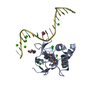
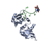
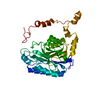
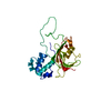
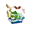
| Title | Crystal structure Analysis of N terminal region containing the dimerization domain and DNA binding domain of HU protein(Histone like protein-DNA binding) from Mycobacterium tuberculosis [H37Ra] | |||||||||
 Components Components | DNA-binding protein HU homolog | |||||||||
 Keywords Keywords | DNA BINDING PROTEIN / DIMERIZATION BY FOUR HELIX BUNDLE INTERACTION / DNA condensation / DNA-binding | |||||||||
| Function / homology |  Function and homology information Function and homology informationbiofilm matrix assembly / : / cellular response to iron ion starvation / DNA protection / nucleoid / chromosome condensation / supercoiled DNA binding / DNA-binding transcription activator activity / ferroxidase / ferroxidase activity ...biofilm matrix assembly / : / cellular response to iron ion starvation / DNA protection / nucleoid / chromosome condensation / supercoiled DNA binding / DNA-binding transcription activator activity / ferroxidase / ferroxidase activity / ferric iron binding / peptidoglycan-based cell wall / iron ion transport / protein-DNA complex / cell wall organization / structural constituent of chromatin / double-stranded DNA binding / damaged DNA binding / intracellular iron ion homeostasis / oxidoreductase activity / transcription cis-regulatory region binding / positive regulation of DNA-templated transcription / DNA binding / extracellular region / plasma membrane / cytosol Similarity search - Function | |||||||||
| Biological species |  Mycobacterium tuberculosis H37Ra (bacteria) Mycobacterium tuberculosis H37Ra (bacteria) | |||||||||
| Method |  X-RAY DIFFRACTION / SYNCHROTRON / MOLECULAR REPLACEMENT / Resolution: 2.04 Å X-RAY DIFFRACTION / SYNCHROTRON / MOLECULAR REPLACEMENT / Resolution: 2.04 Å | |||||||||
 Authors Authors | Bhowmick, T. / Ramagopal, U.A. / Ghosh, S. / Nagaraja, V. / Ramakumar, S. | |||||||||
 Citation Citation | Journal: Nat Commun / Year: 2014 Title: Targeting Mycobacterium tuberculosis nucleoid-associated protein HU with structure-based inhibitors Authors: Bhowmick, T. / Ghosh, S. / Dixit, K. / Ganesan, V. / Ramagopal, U.A. / Dey, D. / Sarma, S.P. / Ramakumar, S. / Nagaraja, V. | |||||||||
| History |
|
- Structure visualization
| Structure viewer | Molecule: MolmilJmol/JSmol |
|---|
- Downloads & links
-Download
| PDBx/mmCIF format | 4pt4.cif.gz | 84.3 KB | Display | PDBx/mmCIF format |
|---|---|---|---|---|
| PDB format | pdb4pt4.ent.gz | 64.1 KB | Display | PDB format |
| PDBx/mmJSON format | 4pt4.json.gz | Tree view | PDBx/mmJSON format | |
| Others |  Other downloads Other downloads |
-Validation report
| Summary document | 4pt4_validation.pdf.gz | 441.8 KB | Display | wwPDB validaton report |
|---|---|---|---|---|
| Full document | 4pt4_full_validation.pdf.gz | 441.9 KB | Display | |
| Data in XML | 4pt4_validation.xml.gz | 9.9 KB | Display | |
| Data in CIF | 4pt4_validation.cif.gz | 13 KB | Display | |
| Arichive directory | https://data.pdbj.org/pub/pdb/validation_reports/pt/4pt4ftp://data.pdbj.org/pub/pdb/validation_reports/pt/4pt4 | HTTPS FTP |
-Related structure data
| Related structure data |  4dkyC 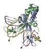 1p71S C: citing same article ( S: Starting model for refinement |
|---|---|
| Similar structure data |


-Links
 PDBj
PDBj
- Assembly
| Deposited unit | 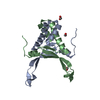
| ||||||||
|---|---|---|---|---|---|---|---|---|---|
| 1 |
| ||||||||
| Unit cell |
|
-Components
| #1: Protein | Mass: 10672.215 Da / Num. of mol.: 2 Fragment: N terminal UNP residues 1-99 (dimerization domain and DNA binding domain) Source method: isolated from a genetically manipulated source Source: (gene. exp.) Mycobacterium tuberculosis H37Ra (bacteria)Gene: hupB, MRA_3015 / Plasmid: pET20b / Production host: #2: Chemical | ChemComp-FMT /   Mass: 46.025 Da / Num. of mol.: 4 / Source method: obtained synthetically / Formula: CH2O2 Mass: 46.025 Da / Num. of mol.: 4 / Source method: obtained synthetically / Formula: CH2O2#3: Water | ChemComp-HOH / |  Mass: 18.015 Da / Num. of mol.: 86 / Source method: isolated from a natural source / Formula: H2O Mass: 18.015 Da / Num. of mol.: 86 / Source method: isolated from a natural source / Formula: H2O |
|---|
-Experimental details
-Experiment
| Experiment | Method: X-RAY DIFFRACTION / Number of used crystals: 1 |
|---|
- Sample preparation
| Crystal | Density Matthews: 1.93 Å3/Da / Density % sol: 36.13 % / Mosaicity: 1.172 ° |
|---|---|
| Crystal grow | Temperature: 293 K / Method: vapor diffusion, hanging drop / pH: 8.2 Details: 3M Sodium formate, 0.1M Tris-Cl, pH 8.2, VAPOR DIFFUSION, HANGING DROP, temperature 293K |
-Data collection
| Diffraction | Mean temperature: 100 K | ||||||||||||||||||||||||||||||||||||||||||||||||||||||||||||||||||
|---|---|---|---|---|---|---|---|---|---|---|---|---|---|---|---|---|---|---|---|---|---|---|---|---|---|---|---|---|---|---|---|---|---|---|---|---|---|---|---|---|---|---|---|---|---|---|---|---|---|---|---|---|---|---|---|---|---|---|---|---|---|---|---|---|---|---|---|
| Diffraction source | Source: SYNCHROTRON / Site: NSLS  / Beamline: X29A / Wavelength: 0.979 Å / Beamline: X29A / Wavelength: 0.979 Å | ||||||||||||||||||||||||||||||||||||||||||||||||||||||||||||||||||
| Detector | Type: ADSC QUANTUM 315 / Detector: CCD / Date: Nov 1, 2007 / Details: mirrors | ||||||||||||||||||||||||||||||||||||||||||||||||||||||||||||||||||
| Radiation | Monochromator: Double Crystal Monochromator (Cryogenically cooled) Protocol: SINGLE WAVELENGTH / Monochromatic (M) / Laue (L): M / Scattering type: x-ray | ||||||||||||||||||||||||||||||||||||||||||||||||||||||||||||||||||
| Radiation wavelength | Wavelength: 0.979 Å / Relative weight: 1 | ||||||||||||||||||||||||||||||||||||||||||||||||||||||||||||||||||
| Reflection | Resolution: 2.04→50 Å / Num. obs: 10304 / % possible obs: 98.7 % / Redundancy: 3.8 % / Rmerge(I) obs: 0.092 / Χ2: 1.078 / Net I/σ(I): 7.5 | ||||||||||||||||||||||||||||||||||||||||||||||||||||||||||||||||||
| Reflection shell | Diffraction-ID: 1 / Rejects: _
|
- Processing
| Software |
| |||||||||||||||||||||||||||||||||||||||||||||||||||||||||||||||||||||||||||
|---|---|---|---|---|---|---|---|---|---|---|---|---|---|---|---|---|---|---|---|---|---|---|---|---|---|---|---|---|---|---|---|---|---|---|---|---|---|---|---|---|---|---|---|---|---|---|---|---|---|---|---|---|---|---|---|---|---|---|---|---|---|---|---|---|---|---|---|---|---|---|---|---|---|---|---|---|
| Refinement | Method to determine structure: MOLECULAR REPLACEMENT Starting model: PDB ENTRY 1P71 Resolution: 2.04→41.42 Å / Cor.coef. Fo:Fc: 0.957 / Cor.coef. Fo:Fc free: 0.923 / WRfactor Rfree: 0.2366 / WRfactor Rwork: 0.1766 / FOM work R set: 0.7719 / SU B: 15.753 / SU ML: 0.183 / SU R Cruickshank DPI: 0.2594 / SU Rfree: 0.2072 / Cross valid method: THROUGHOUT / σ(F): 0 / ESU R: 0.259 / ESU R Free: 0.207 / Stereochemistry target values: MAXIMUM LIKELIHOOD Details: HYDROGENS HAVE BEEN ADDED IN THE RIDING POSITIONS U VALUES
| |||||||||||||||||||||||||||||||||||||||||||||||||||||||||||||||||||||||||||
| Solvent computation | Ion probe radii: 0.8 Å / Shrinkage radii: 0.8 Å / VDW probe radii: 1.2 Å / Solvent model: MASK | |||||||||||||||||||||||||||||||||||||||||||||||||||||||||||||||||||||||||||
| Displacement parameters | Biso max: 82.19 Å2 / Biso mean: 34.753 Å2 / Biso min: 18.77 Å2
| |||||||||||||||||||||||||||||||||||||||||||||||||||||||||||||||||||||||||||
| Refine analyze | Luzzati coordinate error obs: 0.249 Å | |||||||||||||||||||||||||||||||||||||||||||||||||||||||||||||||||||||||||||
| Refinement step | Cycle: LAST / Resolution: 2.04→41.42 Å
| |||||||||||||||||||||||||||||||||||||||||||||||||||||||||||||||||||||||||||
| Refine LS restraints |
| |||||||||||||||||||||||||||||||||||||||||||||||||||||||||||||||||||||||||||
| LS refinement shell | Resolution: 2.037→2.09 Å / Total num. of bins used: 20
| |||||||||||||||||||||||||||||||||||||||||||||||||||||||||||||||||||||||||||
| Refinement TLS params. | Method: refined / Refine-ID: X-RAY DIFFRACTION
| |||||||||||||||||||||||||||||||||||||||||||||||||||||||||||||||||||||||||||
| Refinement TLS group |
|
