 Movie
Movie Controller

[English] 日本語
 Yorodumi
- PDB-4pid: Crystal structure of human adenovirus 2 protease with a weak pyri... -
+ Open data

- Basic information
| Entry | Database: PDB / ID: 4pid | ||||||
|---|---|---|---|---|---|---|---|
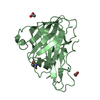
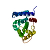
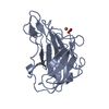
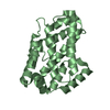
| Title | Crystal structure of human adenovirus 2 protease with a weak pyrimidine nitrile inhibitor | ||||||
 Components Components |
| ||||||
 Keywords Keywords | Hydrolase/Hydrolase Inhibitor / Adenain / cysteine protease / adenovirus / pVIc cofactor / virus maturation / Hydrolase-Hydrolase Inhibitor complex | ||||||
| Function / homology |  Function and homology information Function and homology informationadenain / nuclear capsid assembly / viral procapsid / lysis of host organelle involved in viral entry into host cell / microtubule-dependent intracellular transport of viral material towards nucleus / viral release from host cell / cysteine-type peptidase activity / virion component / viral capsid / host cell ...adenain / nuclear capsid assembly / viral procapsid / lysis of host organelle involved in viral entry into host cell / microtubule-dependent intracellular transport of viral material towards nucleus / viral release from host cell / cysteine-type peptidase activity / virion component / viral capsid / host cell / host cell cytoplasm / cysteine-type endopeptidase activity / host cell nucleus / proteolysis / DNA binding Similarity search - Function | ||||||
| Biological species |   Human adenovirus 2 Human adenovirus 2 | ||||||
| Method |  X-RAY DIFFRACTION / SYNCHROTRON / MOLECULAR REPLACEMENT / Resolution: 1.59 Å X-RAY DIFFRACTION / SYNCHROTRON / MOLECULAR REPLACEMENT / Resolution: 1.59 Å | ||||||
 Authors Authors | Mac Sweeney, A. / Grosche, P. / Ellis, D. / Combrink, K. / Erbel, P. / Hughes, N. / Sirockin, F. / Melkko, S. / Bernardi, A. / Ramage, P. ...Mac Sweeney, A. / Grosche, P. / Ellis, D. / Combrink, K. / Erbel, P. / Hughes, N. / Sirockin, F. / Melkko, S. / Bernardi, A. / Ramage, P. / Jarousse, N. / Altmann, E. | ||||||
 Citation Citation | Journal: Acs Med.Chem.Lett. / Year: 2014 Title: Discovery and structure-based optimization of adenain inhibitors. Authors: Mac Sweeney, A. / Grosche, P. / Ellis, D. / Combrink, K. / Erbel, P. / Hughes, N. / Sirockin, F. / Melkko, S. / Bernardi, A. / Ramage, P. / Jarousse, N. / Altmann, E. | ||||||
| History |
|
- Structure visualization
| Structure viewer | Molecule: MolmilJmol/JSmol |
|---|
- Downloads & links
-Download
| PDBx/mmCIF format | 4pid.cif.gz | 62.9 KB | Display | PDBx/mmCIF format |
|---|---|---|---|---|
| PDB format | pdb4pid.ent.gz | 44.5 KB | Display | PDB format |
| PDBx/mmJSON format | 4pid.json.gz | Tree view | PDBx/mmJSON format | |
| Others |  Other downloads Other downloads |
-Validation report
| Arichive directory | https://data.pdbj.org/pub/pdb/validation_reports/pi/4pidftp://data.pdbj.org/pub/pdb/validation_reports/pi/4pid | HTTPS FTP |
|---|
-Related structure data
| Related structure data | 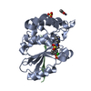 4pieC 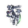 4piqC 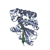 4pisC 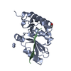 1nlnS S: Starting model for refinement C: citing same article ( |
|---|---|
| Similar structure data |

-Links
 PDBj
PDBj
- Assembly
| Deposited unit | 
| ||||||||
|---|---|---|---|---|---|---|---|---|---|
| 1 |
| ||||||||
| Unit cell |
|
-Components
| #1: Protein | Mass: 23114.350 Da / Num. of mol.: 1 Source method: isolated from a genetically manipulated source Source: (gene. exp.) Human adenovirus 2 / Gene: L3 / Production host:  |
|---|---|
| #2: Protein/peptide | Mass: 1353.640 Da / Num. of mol.: 1 / Fragment: UNP residues 240-250 / Source method: obtained synthetically / Source: (synth.) Human adenovirus 2 / References: UniProt: P03274 |
| #3: Chemical | ChemComp-ACT /   Mass: 59.044 Da / Num. of mol.: 1 / Source method: obtained synthetically / Formula: C2H3O2 Mass: 59.044 Da / Num. of mol.: 1 / Source method: obtained synthetically / Formula: C2H3O2 |
| #4: Chemical | ChemComp-2UQ /   Mass: 240.261 Da / Num. of mol.: 1 / Source method: obtained synthetically / Formula: C13H12N4O Mass: 240.261 Da / Num. of mol.: 1 / Source method: obtained synthetically / Formula: C13H12N4O |
| #5: Water | ChemComp-HOH /  Mass: 18.015 Da / Num. of mol.: 151 / Source method: isolated from a natural source / Formula: H2O Mass: 18.015 Da / Num. of mol.: 151 / Source method: isolated from a natural source / Formula: H2O |
| Has protein modification | Y |
-Experimental details
-Experiment
| Experiment | Method: X-RAY DIFFRACTION / Number of used crystals: 1 |
|---|
- Sample preparation
| Crystal | Density Matthews: 3.78 Å3/Da / Density % sol: 67.44 % |
|---|---|
| Crystal grow | Temperature: 293 K / Method: vapor diffusion, sitting drop / pH: 7.5 Details: 0.8 M sodium/potassium tartrate, 0.1 M HEPES pH 7.5 |
-Data collection
| Diffraction | Mean temperature: 100 K | ||||||||||||||||||||||||||||||||||||||||||||||||||||||||||||||||||||||||||||||||||||||||||||||||||||||||||||||||||||||||||||||||||||||||||||||||||||||||||||||||||||||||||||||||||||||||||||||||||||||||||||||||||
|---|---|---|---|---|---|---|---|---|---|---|---|---|---|---|---|---|---|---|---|---|---|---|---|---|---|---|---|---|---|---|---|---|---|---|---|---|---|---|---|---|---|---|---|---|---|---|---|---|---|---|---|---|---|---|---|---|---|---|---|---|---|---|---|---|---|---|---|---|---|---|---|---|---|---|---|---|---|---|---|---|---|---|---|---|---|---|---|---|---|---|---|---|---|---|---|---|---|---|---|---|---|---|---|---|---|---|---|---|---|---|---|---|---|---|---|---|---|---|---|---|---|---|---|---|---|---|---|---|---|---|---|---|---|---|---|---|---|---|---|---|---|---|---|---|---|---|---|---|---|---|---|---|---|---|---|---|---|---|---|---|---|---|---|---|---|---|---|---|---|---|---|---|---|---|---|---|---|---|---|---|---|---|---|---|---|---|---|---|---|---|---|---|---|---|---|---|---|---|---|---|---|---|---|---|---|---|---|---|---|---|---|
| Diffraction source | Source: SYNCHROTRON / Site: SLS  / Beamline: X10SA / Wavelength: 1 Å / Beamline: X10SA / Wavelength: 1 Å | ||||||||||||||||||||||||||||||||||||||||||||||||||||||||||||||||||||||||||||||||||||||||||||||||||||||||||||||||||||||||||||||||||||||||||||||||||||||||||||||||||||||||||||||||||||||||||||||||||||||||||||||||||
| Detector | Type: PSI PILATUS 6M / Detector: PIXEL / Date: Nov 20, 2011 | ||||||||||||||||||||||||||||||||||||||||||||||||||||||||||||||||||||||||||||||||||||||||||||||||||||||||||||||||||||||||||||||||||||||||||||||||||||||||||||||||||||||||||||||||||||||||||||||||||||||||||||||||||
| Radiation | Protocol: SINGLE WAVELENGTH / Monochromatic (M) / Laue (L): M / Scattering type: x-ray | ||||||||||||||||||||||||||||||||||||||||||||||||||||||||||||||||||||||||||||||||||||||||||||||||||||||||||||||||||||||||||||||||||||||||||||||||||||||||||||||||||||||||||||||||||||||||||||||||||||||||||||||||||
| Radiation wavelength | Wavelength: 1 Å / Relative weight: 1 | ||||||||||||||||||||||||||||||||||||||||||||||||||||||||||||||||||||||||||||||||||||||||||||||||||||||||||||||||||||||||||||||||||||||||||||||||||||||||||||||||||||||||||||||||||||||||||||||||||||||||||||||||||
| Reflection | Resolution: 1.59→97.7 Å / Num. obs: 49317 / % possible obs: 100 % / Observed criterion σ(I): -3 / Redundancy: 10 % / Biso Wilson estimate: 24.337 Å2 / Rmerge F obs: 0.059 / Rmerge(I) obs: 0.063 / Rrim(I) all: 0.066 / Χ2: 1.009 / Net I/σ(I): 22.95 / Num. measured all: 495405 | ||||||||||||||||||||||||||||||||||||||||||||||||||||||||||||||||||||||||||||||||||||||||||||||||||||||||||||||||||||||||||||||||||||||||||||||||||||||||||||||||||||||||||||||||||||||||||||||||||||||||||||||||||
| Reflection shell | Diffraction-ID: 1 / Rejects: _
|
- Processing
| Software |
| |||||||||||||||||||||||||||||||||||||||||||||||||||||||||||||||||
|---|---|---|---|---|---|---|---|---|---|---|---|---|---|---|---|---|---|---|---|---|---|---|---|---|---|---|---|---|---|---|---|---|---|---|---|---|---|---|---|---|---|---|---|---|---|---|---|---|---|---|---|---|---|---|---|---|---|---|---|---|---|---|---|---|---|---|
| Refinement | Method to determine structure: MOLECULAR REPLACEMENT Starting model: PDB entry 1NLN Resolution: 1.59→48.84 Å / Cor.coef. Fo:Fc: 0.967 / Cor.coef. Fo:Fc free: 0.957 / SU B: 0.973 / SU ML: 0.035 / Cross valid method: THROUGHOUT / σ(F): 0 / ESU R: 0.059 / ESU R Free: 0.062 / Stereochemistry target values: MAXIMUM LIKELIHOOD Details: HYDROGENS HAVE BEEN ADDED IN THE RIDING POSITIONS U VALUES : REFINED INDIVIDUALLY
| |||||||||||||||||||||||||||||||||||||||||||||||||||||||||||||||||
| Solvent computation | Ion probe radii: 0.8 Å / Shrinkage radii: 0.8 Å / VDW probe radii: 1.4 Å / Solvent model: MASK | |||||||||||||||||||||||||||||||||||||||||||||||||||||||||||||||||
| Displacement parameters | Biso max: 58.91 Å2 / Biso mean: 18.571 Å2 / Biso min: 5.61 Å2
| |||||||||||||||||||||||||||||||||||||||||||||||||||||||||||||||||
| Refinement step | Cycle: final / Resolution: 1.59→48.84 Å
| |||||||||||||||||||||||||||||||||||||||||||||||||||||||||||||||||
| Refine LS restraints |
| |||||||||||||||||||||||||||||||||||||||||||||||||||||||||||||||||
| LS refinement shell | Resolution: 1.59→1.631 Å / Total num. of bins used: 20
|
