| Entry | Database: PDB / ID: 4p8u
|
|---|
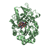
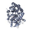
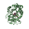
| Title | The crystal structures of YKL-39 in the absence of chitooligosaccharides was solved to resolutions of 2.4 angstrom |
|---|
 Components Components | Chitinase-3-like protein 2 |
|---|
 Keywords Keywords | SUGAR BINDING PROTEIN / chitinase 3-like protein 2 / human YKL-39 / family-18 chitinase |
|---|
| Function / homology |  Function and homology information Function and homology information
chitin catabolic process / chitin binding / carbohydrate binding / carbohydrate metabolic process / hydrolase activity / : / extracellular regionSimilarity search - Function Chitinase A; domain 3 - #10 / : / Chitinase insertion domain superfamily / Chitinase II / Glyco_18 / Glycosyl hydrolases family 18 / Glycosyl hydrolases family 18 (GH18) domain profile. / Glycoside hydrolase family 18, catalytic domain / Chitinase A; domain 3 / Glycosidases ...Chitinase A; domain 3 - #10 / : / Chitinase insertion domain superfamily / Chitinase II / Glyco_18 / Glycosyl hydrolases family 18 / Glycosyl hydrolases family 18 (GH18) domain profile. / Glycoside hydrolase family 18, catalytic domain / Chitinase A; domain 3 / Glycosidases / Glycoside hydrolase superfamily / TIM Barrel / Alpha-Beta Barrel / Roll / Alpha BetaSimilarity search - Domain/homology |
|---|
| Biological species |  Homo sapiens (human) Homo sapiens (human) |
|---|
| Method |  X-RAY DIFFRACTION / SYNCHROTRON / MOLECULAR REPLACEMENT / Resolution: 2.4 Å X-RAY DIFFRACTION / SYNCHROTRON / MOLECULAR REPLACEMENT / Resolution: 2.4 Å |
|---|
 Authors Authors | Suginta, W. / Ranok, A. / Robinson, R.C. / Wongsantichon, J. |
|---|
| Funding support |  Thailand, 1items Thailand, 1items | Organization | Grant number | Country |
|---|
| the Office of the Higher Education Commission | CHE500307 | Thailand |
|
|---|
 Citation Citation | Journal: J.Biol.Chem. / Year: 2015
Title: Structural and Thermodynamic Insights into Chitooligosaccharide Binding to Human Cartilage Chitinase 3-like Protein 2 (CHI3L2 or YKL-39).
Authors: Ranok, A. / Wongsantichon, J. / Robinson, R.C. / Suginta, W. |
|---|
| History | | Deposition | Apr 1, 2014 | Deposition site: RCSB / Processing site: RCSB |
|---|
| Revision 1.0 | Oct 1, 2014 | Provider: repository / Type: Initial release |
|---|
| Revision 1.1 | Dec 17, 2014 | Group: Database references |
|---|
| Revision 1.2 | Feb 4, 2015 | Group: Derived calculations |
|---|
| Revision 1.3 | Feb 11, 2015 | Group: Database references |
|---|
| Revision 1.4 | Dec 27, 2023 | Group: Data collection / Database references ...Data collection / Database references / Derived calculations / Other / Refinement description / Source and taxonomy / Structure summary
Category: chem_comp_atom / chem_comp_bond ...chem_comp_atom / chem_comp_bond / citation / database_2 / entity_src_gen / pdbx_database_status / pdbx_struct_assembly / pdbx_struct_oper_list / refine_hist / struct_keywords
Item: _citation.journal_id_CSD / _database_2.pdbx_DOI ..._citation.journal_id_CSD / _database_2.pdbx_DOI / _database_2.pdbx_database_accession / _entity_src_gen.pdbx_alt_source_flag / _pdbx_database_status.pdb_format_compatible / _pdbx_struct_assembly.oligomeric_details / _pdbx_struct_oper_list.symmetry_operation / _refine_hist.number_atoms_solvent / _refine_hist.pdbx_number_atoms_ligand / _refine_hist.pdbx_number_atoms_nucleic_acid / _refine_hist.pdbx_number_atoms_protein / _struct_keywords.text |
|---|
| Revision 1.5 | Nov 20, 2024 | Group: Structure summary / Category: pdbx_entry_details / pdbx_modification_feature |
|---|
|
|---|
 Movie
Movie Controller

 Yorodumi
 Open data
 Basic information
 Structure visualization
 Downloads & links
 Other downloads


 PDBj
PDBj Assembly


 Mass: 96.063 Da / Num. of mol.: 2 / Source method: obtained synthetically / Formula: SO4
 Mass: 106.120 Da / Num. of mol.: 1 / Source method: obtained synthetically / Formula: C4H10O3
 Mass: 18.015 Da / Num. of mol.: 168 / Source method: isolated from a natural source / Formula: H2O
 Sample preparation
 / Beamline: BL13B1 / Wavelength: 1 Å
 Processing