- PDB-5z4v: Crystal structure of the sheep signalling glycoprotein (SPS-40) c... -
+
Open data
ID or keywords:
Loading...
-
Basic information
Entry
Database: PDB / ID: 5z4v
Title
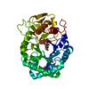
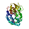
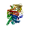
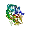
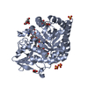
Crystal structure of the sheep signalling glycoprotein (SPS-40) complex with 2-methyl-2-4-pentanediol at 1.65A resolution reveals specific binding characteristics of SPS-40
Components
Chitinase-3-like protein 1
Keywords
SIGNALING PROTEIN
Function / homology
Function and homology information
response to interleukin-6 / activation of NF-kappaB-inducing kinase activity / positive regulation of peptidyl-threonine phosphorylation / chitin catabolic process / chitin binding / response to tumor necrosis factor / response to mechanical stimulus / lung development / response to interleukin-1 / positive regulation of interleukin-8 production ...response to interleukin-6 / activation of NF-kappaB-inducing kinase activity / positive regulation of peptidyl-threonine phosphorylation / chitin catabolic process / chitin binding / response to tumor necrosis factor / response to mechanical stimulus / lung development / response to interleukin-1 / positive regulation of interleukin-8 production / cellular response to tumor necrosis factor / positive regulation of angiogenesis / carbohydrate binding / carbohydrate metabolic process / positive regulation of ERK1 and ERK2 cascade / positive regulation of phosphatidylinositol 3-kinase/protein kinase B signal transduction / inflammatory response / apoptotic process / perinuclear region of cytoplasm / endoplasmic reticulum / : / cytoplasm Similarity search - Function
: / Chitinase insertion domain superfamily / Chitinase II / Glyco_18 / Glycosyl hydrolases family 18 / Glycosyl hydrolases family 18 (GH18) domain profile. / Glycoside hydrolase family 18, catalytic domain / Glycoside hydrolase superfamily Similarity search - Domain/homology
Mass: 18.015 Da / Num. of mol.: 599 / Source method: isolated from a natural source / Formula: H2O
Has protein modification
Y
Sequence details
CONFLICTS ARE OBSERVED BETWEEN THE SEQUENCE IN THIS STRUCTURE AND DATABASE UNIPROTKB Q6TMG6 (CH3L1_ ...CONFLICTS ARE OBSERVED BETWEEN THE SEQUENCE IN THIS STRUCTURE AND DATABASE UNIPROTKB Q6TMG6 (CH3L1_SHEEP) THE AUTHOR EXPECTS SEQUENCE IN THIS STRUCTURE TO BE AN ISOFORM. RESIDUE NUMBER 211 IS SIMPLY SKIPPED.
-
Experimental details
-
Experiment
Experiment
Method: X-RAY DIFFRACTION / Number of used crystals: 1
-
Sample preparation
Crystal
Density Matthews: 2.62 Å3/Da / Density % sol: 53.14 %
Crystal grow
Temperature: 298 K / Method: vapor diffusion, hanging drop / pH: 7.8 Details: Tri-HCl, 20% Ethanol, pH 7.8, VAPOR DIFFUSION, HANGING DROP, temperature 298K PH range: 5-8
Resolution: 1.65→27.711 Å / Cor.coef. Fo:Fc: 0.977 / Cor.coef. Fo:Fc free: 0.959 / SU B: 3.49 / SU ML: 0.053 / Cross valid method: THROUGHOUT / ESU R: 0.071 / ESU R Free: 0.082 / Stereochemistry target values: MAXIMUM LIKELIHOOD / Details: HYDROGENS HAVE BEEN ADDED IN THE RIDING POSITIONS
Rfactor
Num. reflection
% reflection
Selection details
Rfree
0.1876
1075
2.1 %
RANDOM
Rwork
0.13756
-
-
-
obs
0.1385
50989
99.88 %
-
Solvent computation
Ion probe radii: 0.8 Å / Shrinkage radii: 0.8 Å / VDW probe radii: 1.2 Å / Solvent model: MASK
 Movie
Movie Controller

 Yorodumi
 Open data
 Basic information
 Components
 Keywords
 Function and homology information
 X-RAY DIFFRACTION /
  Authors
 Citation
 Structure visualization
 Downloads & links
 Other downloads
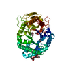


 PDBj
PDBj Assembly

 Type: D-saccharide, beta linking / Mass: 221.208 Da / Num. of mol.: 1
 Mass: 118.174 Da / Num. of mol.: 2 / Source method: obtained synthetically / Formula: C6H14O2 / Comment: precipitant*YM
 Mass: 18.015 Da / Num. of mol.: 599 / Source method: isolated from a natural source / Formula: H2O
 Sample preparation
 / Beamline: X13 / Wavelength: 0.81 Å
 Processing