 Movie
Movie Controller


+ Open data

- Basic information
| Entry | Database: PDB / ID: 4p37 | ||||||
|---|---|---|---|---|---|---|---|
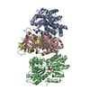
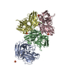
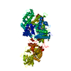
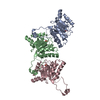
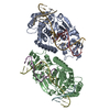
| Title | Crystal structure of the Megavirus polyadenylate synthase | ||||||
 Components Components | (Putative poly(A) polymerase catalytic ...) x 2 | ||||||
 Keywords Keywords | TRANSFERASE / PolyA polymerase | ||||||
| Function / homology |  Function and homology information Function and homology informationpolynucleotide adenylyltransferase / virion component / mRNA processing / transferase activity Similarity search - Function | ||||||
| Biological species |  Megavirus chiliensis Megavirus chiliensis | ||||||
| Method |  X-RAY DIFFRACTION / SYNCHROTRON / MAD / Resolution: 2.24 Å X-RAY DIFFRACTION / SYNCHROTRON / MAD / Resolution: 2.24 Å | ||||||
 Authors Authors | Priet, S. / Lartigue, A. / Claverie, J.M. / Abergel, C. | ||||||
 Citation Citation | Journal: Nucleic Acids Res. / Year: 2015 Title: mRNA maturation in giant viruses: variation on a theme. Authors: Priet, S. / Lartigue, A. / Debart, F. / Claverie, J.M. / Abergel, C. | ||||||
| History |
|
- Structure visualization
| Structure viewer | Molecule: MolmilJmol/JSmol |
|---|
- Downloads & links
-Download
| PDBx/mmCIF format | 4p37.cif.gz | 237.4 KB | Display | PDBx/mmCIF format |
|---|---|---|---|---|
| PDB format | pdb4p37.ent.gz | 188.8 KB | Display | PDB format |
| PDBx/mmJSON format | 4p37.json.gz | Tree view | PDBx/mmJSON format | |
| Others |  Other downloads Other downloads |
-Validation report
| Arichive directory | https://data.pdbj.org/pub/pdb/validation_reports/p3/4p37ftp://data.pdbj.org/pub/pdb/validation_reports/p3/4p37 | HTTPS FTP |
|---|
-Related structure data


-Links
 PDBj
PDBj
- Assembly
| Deposited unit | 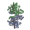
| ||||||||
|---|---|---|---|---|---|---|---|---|---|
| 1 |
| ||||||||
| Unit cell |
| ||||||||
| Details | Gel filtration confirms the dimerization of the protein in solution |
-Components
-Putative poly(A) polymerase catalytic ... , 2 types, 2 molecules AB
| #1: Protein | Mass: 63666.695 Da / Num. of mol.: 1 Source method: isolated from a genetically manipulated source Source: (gene. exp.) Megavirus chiliensis / Gene: mg561 / Plasmid: modified pETDuet / Production host:  |
|---|---|
| #2: Protein | Mass: 63707.727 Da / Num. of mol.: 1 Source method: isolated from a genetically manipulated source Source: (gene. exp.) Megavirus chiliensis / Gene: mg561 / Plasmid: modified pETDuet / Production host: |
-Non-polymers , 4 types, 592 molecules 


| #3: Chemical | ChemComp-PEG /  Mass: 106.120 Da / Num. of mol.: 10 / Source method: obtained synthetically / Formula: C4H10O3 Mass: 106.120 Da / Num. of mol.: 10 / Source method: obtained synthetically / Formula: C4H10O3#4: Chemical | ChemComp-TRS / |  Mass: 122.143 Da / Num. of mol.: 1 / Source method: obtained synthetically / Formula: C4H12NO3 / Comment: pH buffer*YM Mass: 122.143 Da / Num. of mol.: 1 / Source method: obtained synthetically / Formula: C4H12NO3 / Comment: pH buffer*YM#5: Chemical |  Mass: 62.068 Da / Num. of mol.: 3 / Source method: obtained synthetically / Formula: C2H6O2 Mass: 62.068 Da / Num. of mol.: 3 / Source method: obtained synthetically / Formula: C2H6O2#6: Water | ChemComp-HOH / | Mass: 18.015 Da / Num. of mol.: 578 / Source method: isolated from a natural source / Formula: H2O |
|---|
-Details
| Has protein modification | Y |
|---|
-Experimental details
-Experiment
| Experiment | Method: X-RAY DIFFRACTION |
|---|
- Sample preparation
| Crystal | Density Matthews: 2.55 Å3/Da / Density % sol: 51.85 % |
|---|---|
| Crystal grow | Temperature: 293 K / Method: vapor diffusion, hanging drop / pH: 6.8 / Details: 0.1M Tris-HCl, PEG 200 |
-Data collection
| Diffraction | Mean temperature: 105 K | ||||||||||||
|---|---|---|---|---|---|---|---|---|---|---|---|---|---|
| Diffraction source | Source: SYNCHROTRON / Site: ESRF  / Beamline: ID29 / Wavelength: 0.9793, 0.9795, 0.9770 / Beamline: ID29 / Wavelength: 0.9793, 0.9795, 0.9770 | ||||||||||||
| Detector | Type: DECTRIS PILATUS 6M / Detector: PIXEL / Date: Oct 21, 2011 | ||||||||||||
| Radiation | Protocol: MAD / Monochromatic (M) / Laue (L): M / Scattering type: x-ray | ||||||||||||
| Radiation wavelength |
| ||||||||||||
| Reflection | Resolution: 2.24→51.3 Å / Num. obs: 62550 / % possible obs: 99 % / Redundancy: 3.9 % / Biso Wilson estimate: 49.16 Å2 / Rmerge(I) obs: 0.069 / Net I/σ(I): 6.7 | ||||||||||||
| Reflection shell | Resolution: 2.24→2.36 Å / Redundancy: 4.1 % / Mean I/σ(I) obs: 1.5 / % possible all: 99 |
- Processing
| Software |
| ||||||||||||||||||||||||||||||||||||||||||||||||||||||||||||||||||||||||||||||||||||||||||||||||||||||||||||||||||
|---|---|---|---|---|---|---|---|---|---|---|---|---|---|---|---|---|---|---|---|---|---|---|---|---|---|---|---|---|---|---|---|---|---|---|---|---|---|---|---|---|---|---|---|---|---|---|---|---|---|---|---|---|---|---|---|---|---|---|---|---|---|---|---|---|---|---|---|---|---|---|---|---|---|---|---|---|---|---|---|---|---|---|---|---|---|---|---|---|---|---|---|---|---|---|---|---|---|---|---|---|---|---|---|---|---|---|---|---|---|---|---|---|---|---|---|
| Refinement | Method to determine structure: MAD / Resolution: 2.24→49.38 Å / Cor.coef. Fo:Fc: 0.9351 / Cor.coef. Fo:Fc free: 0.9181 / SU R Cruickshank DPI: 0.247 / Cross valid method: THROUGHOUT / σ(F): 0 / SU R Blow DPI: 0.255 / SU Rfree Blow DPI: 0.184 / SU Rfree Cruickshank DPI: 0.183
| ||||||||||||||||||||||||||||||||||||||||||||||||||||||||||||||||||||||||||||||||||||||||||||||||||||||||||||||||||
| Displacement parameters | Biso mean: 55.14 Å2
| ||||||||||||||||||||||||||||||||||||||||||||||||||||||||||||||||||||||||||||||||||||||||||||||||||||||||||||||||||
| Refine analyze | Luzzati coordinate error obs: 0.276 Å | ||||||||||||||||||||||||||||||||||||||||||||||||||||||||||||||||||||||||||||||||||||||||||||||||||||||||||||||||||
| Refinement step | Cycle: LAST / Resolution: 2.24→49.38 Å
| ||||||||||||||||||||||||||||||||||||||||||||||||||||||||||||||||||||||||||||||||||||||||||||||||||||||||||||||||||
| Refine LS restraints |
| ||||||||||||||||||||||||||||||||||||||||||||||||||||||||||||||||||||||||||||||||||||||||||||||||||||||||||||||||||
| LS refinement shell | Resolution: 2.24→2.3 Å / Total num. of bins used: 20
|
