 Movie
Movie Controller

[English] 日本語
 Yorodumi
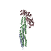
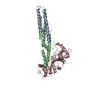
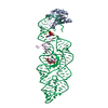
- PDB-4p2y: Crystal structure of the human RAGE ectodomain (fragment VC1C2) i... -
+ Open data

- Basic information
| Entry | Database: PDB / ID: 4p2y | ||||||
|---|---|---|---|---|---|---|---|
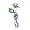
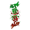
| Title | Crystal structure of the human RAGE ectodomain (fragment VC1C2) in complex with mouse S100A6 | ||||||
 Components Components |
| ||||||
 Keywords Keywords | SIGNALING PROTEIN / signaling complex / pattern recognition receptor / dimerization / EF-hand calcium binding protein | ||||||
| Function / homology |  Function and homology information Function and homology informationregulation of CD4-positive, alpha-beta T cell activation / advanced glycation end-product receptor activity / negative regulation of blood circulation / positive regulation of endothelin production / positive regulation of DNA-templated DNA replication / glucose mediated signaling pathway / positive regulation of monocyte extravasation / regulation of T cell mediated cytotoxicity / positive regulation of dendritic cell differentiation / negative regulation of long-term synaptic depression ...regulation of CD4-positive, alpha-beta T cell activation / advanced glycation end-product receptor activity / negative regulation of blood circulation / positive regulation of endothelin production / positive regulation of DNA-templated DNA replication / glucose mediated signaling pathway / positive regulation of monocyte extravasation / regulation of T cell mediated cytotoxicity / positive regulation of dendritic cell differentiation / negative regulation of long-term synaptic depression / regulation of p38MAPK cascade / monoatomic ion transmembrane transporter activity / regulation of non-canonical NF-kappaB signal transduction / positive regulation of amyloid precursor protein catabolic process / transcytosis / induction of positive chemotaxis / S100 protein binding / positive regulation of monocyte chemotactic protein-1 production / regulation of long-term synaptic potentiation / positive regulation of p38MAPK cascade / positive regulation of heterotypic cell-cell adhesion / positive regulation of activated T cell proliferation / negative regulation of connective tissue replacement involved in inflammatory response wound healing / protein localization to membrane / scavenger receptor activity / regulation of spontaneous synaptic transmission / negative regulation of interleukin-10 production / positive regulation of double-strand break repair / tropomyosin binding / response to amyloid-beta / laminin receptor activity / TRAF6 mediated NF-kB activation / negative regulation of long-term synaptic potentiation / Advanced glycosylation endproduct receptor signaling / phagocytosis / transport across blood-brain barrier / phagocytic cup / positive regulation of chemokine production / ruffle / positive regulation of interleukin-12 production / astrocyte activation / : / positive regulation of interleukin-1 beta production / microglial cell activation / positive regulation of non-canonical NF-kappaB signal transduction / TAK1-dependent IKK and NF-kappa-B activation / response to wounding / regulation of synaptic plasticity / positive regulation of interleukin-6 production / positive regulation of JNK cascade / fibrillar center / cellular response to amyloid-beta / cytoplasmic side of plasma membrane / neuron projection development / positive regulation of tumor necrosis factor production / calcium-dependent protein binding / cell junction / transmembrane signaling receptor activity / nuclear envelope / amyloid-beta binding / extracellular matrix / signaling receptor activity / regulation of inflammatory response / histone binding / molecular adaptor activity / learning or memory / response to hypoxia / early endosome / positive regulation of ERK1 and ERK2 cascade / DNA replication / cell surface receptor signaling pathway / apical plasma membrane / postsynapse / inflammatory response / DNA repair / calcium ion binding / protein-containing complex binding / perinuclear region of cytoplasm / cell surface / protein homodimerization activity / DNA binding / RNA binding / extracellular region / zinc ion binding / identical protein binding / nucleus / plasma membrane / cytoplasm / cytosol Similarity search - Function | ||||||
| Biological species |  Homo sapiens (human) Homo sapiens (human) | ||||||
| Method |  X-RAY DIFFRACTION / SYNCHROTRON / MOLECULAR REPLACEMENT / molecular replacement / Resolution: 2.3 Å X-RAY DIFFRACTION / SYNCHROTRON / MOLECULAR REPLACEMENT / molecular replacement / Resolution: 2.3 Å | ||||||
 Authors Authors | Yatime, L. / Andersen, G.R. | ||||||
 Citation Citation | Journal: Structure / Year: 2016 Title: The Structure of the RAGE:S100A6 Complex Reveals a Unique Mode of Homodimerization for S100 Proteins. Authors: Yatime, L. / Betzer, C. / Jensen, R.K. / Mortensen, S. / Jensen, P.H. / Andersen, G.R. | ||||||
| History |
|
- Structure visualization
| Structure viewer | Molecule: MolmilJmol/JSmol |
|---|
- Downloads & links
-Download
| PDBx/mmCIF format | 4p2y.cif.gz | 177.7 KB | Display | PDBx/mmCIF format |
|---|---|---|---|---|
| PDB format | pdb4p2y.ent.gz | 138.1 KB | Display | PDB format |
| PDBx/mmJSON format | 4p2y.json.gz | Tree view | PDBx/mmJSON format | |
| Others |  Other downloads Other downloads |
-Validation report
| Arichive directory | https://data.pdbj.org/pub/pdb/validation_reports/p2/4p2yftp://data.pdbj.org/pub/pdb/validation_reports/p2/4p2y | HTTPS FTP |
|---|
-Related structure data
| Related structure data | 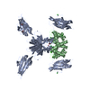 4ybhC  1k96S  4lp5S S: Starting model for refinement C: citing same article ( |
|---|---|
| Similar structure data |


-Links
 PDBj
PDBj


- Assembly
| Deposited unit | 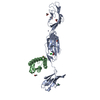
| ||||||||
|---|---|---|---|---|---|---|---|---|---|
| 1 |
| ||||||||
| 2 | 
| ||||||||
| 3 | 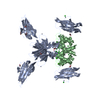
| ||||||||
| Unit cell |
| ||||||||
| Details | The biological assembly is a heterotetramer generated from the heterodimer in the asymmetric unit by the operation: -x, y, -z. |
-Components
-Protein , 2 types, 2 molecules AB
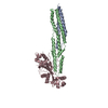
| #1: Protein | Mass: 32680.297 Da / Num. of mol.: 1 Fragment: V, C1 and C2 domains (VC1C2 module), full-length ectodomain, UNP residues 23-323 Source method: isolated from a genetically manipulated source Source: (gene. exp.) Homo sapiens (human) / Gene: AGER, RAGE / Plasmid: pETM11 / Production host:  |
|---|---|
| #2: Protein | Mass: 10192.740 Da / Num. of mol.: 1 Source method: isolated from a genetically manipulated source Source: (gene. exp.) |
-Non-polymers , 5 types, 207 molecules 


| #3: Chemical | ChemComp-ZN /  Mass: 65.409 Da / Num. of mol.: 6 / Source method: obtained synthetically / Formula: Zn Mass: 65.409 Da / Num. of mol.: 6 / Source method: obtained synthetically / Formula: Zn#4: Chemical | ChemComp-CL /  Mass: 35.453 Da / Num. of mol.: 6 / Source method: obtained synthetically / Formula: Cl Mass: 35.453 Da / Num. of mol.: 6 / Source method: obtained synthetically / Formula: Cl#5: Chemical | ChemComp-ACT /  Mass: 59.044 Da / Num. of mol.: 7 / Source method: obtained synthetically / Formula: C2H3O2 Mass: 59.044 Da / Num. of mol.: 7 / Source method: obtained synthetically / Formula: C2H3O2#6: Chemical |  Mass: 40.078 Da / Num. of mol.: 2 / Source method: obtained synthetically / Formula: Ca Mass: 40.078 Da / Num. of mol.: 2 / Source method: obtained synthetically / Formula: Ca#7: Water | ChemComp-HOH / | Mass: 18.015 Da / Num. of mol.: 186 / Source method: isolated from a natural source / Formula: H2O |
|---|
-Details
| Has protein modification | Y |
|---|
-Experimental details
-Experiment
| Experiment | Method: X-RAY DIFFRACTION / Number of used crystals: 1 |
|---|
- Sample preparation
| Crystal | Density Matthews: 3.61 Å3/Da / Density % sol: 65.9 % |
|---|---|
| Crystal grow | Temperature: 277 K / Method: vapor diffusion, hanging drop / pH: 6.5 Details: 0.2 M Zn acetate, 0.1 M Na cacodylate pH 6.5, 10% isopropanol |
-Data collection
| Diffraction | Mean temperature: 100 K | ||||||||||||||||||||||||||||||||||||||||||||||||||||||||||||||||||||||||||||||||||||||||||||||||||||||||||||||||||||||||||||||||||||||||||||||||||||||||||||||||||||||||||||||||||||||||||||||||||||||||||||||||||||||||||||
|---|---|---|---|---|---|---|---|---|---|---|---|---|---|---|---|---|---|---|---|---|---|---|---|---|---|---|---|---|---|---|---|---|---|---|---|---|---|---|---|---|---|---|---|---|---|---|---|---|---|---|---|---|---|---|---|---|---|---|---|---|---|---|---|---|---|---|---|---|---|---|---|---|---|---|---|---|---|---|---|---|---|---|---|---|---|---|---|---|---|---|---|---|---|---|---|---|---|---|---|---|---|---|---|---|---|---|---|---|---|---|---|---|---|---|---|---|---|---|---|---|---|---|---|---|---|---|---|---|---|---|---|---|---|---|---|---|---|---|---|---|---|---|---|---|---|---|---|---|---|---|---|---|---|---|---|---|---|---|---|---|---|---|---|---|---|---|---|---|---|---|---|---|---|---|---|---|---|---|---|---|---|---|---|---|---|---|---|---|---|---|---|---|---|---|---|---|---|---|---|---|---|---|---|---|---|---|---|---|---|---|---|---|---|---|---|---|---|---|---|---|---|
| Diffraction source | Source: SYNCHROTRON / Site: MAX II  / Beamline: I911-2 / Wavelength: 1.041 Å / Beamline: I911-2 / Wavelength: 1.041 Å | ||||||||||||||||||||||||||||||||||||||||||||||||||||||||||||||||||||||||||||||||||||||||||||||||||||||||||||||||||||||||||||||||||||||||||||||||||||||||||||||||||||||||||||||||||||||||||||||||||||||||||||||||||||||||||||
| Detector | Type: MAR CCD 165 mm / Detector: CCD / Date: Feb 19, 2012 | ||||||||||||||||||||||||||||||||||||||||||||||||||||||||||||||||||||||||||||||||||||||||||||||||||||||||||||||||||||||||||||||||||||||||||||||||||||||||||||||||||||||||||||||||||||||||||||||||||||||||||||||||||||||||||||
| Radiation | Monochromator: Si(111) / Protocol: SINGLE WAVELENGTH / Scattering type: x-ray | ||||||||||||||||||||||||||||||||||||||||||||||||||||||||||||||||||||||||||||||||||||||||||||||||||||||||||||||||||||||||||||||||||||||||||||||||||||||||||||||||||||||||||||||||||||||||||||||||||||||||||||||||||||||||||||
| Radiation wavelength | Wavelength: 1.041 Å / Relative weight: 1 | ||||||||||||||||||||||||||||||||||||||||||||||||||||||||||||||||||||||||||||||||||||||||||||||||||||||||||||||||||||||||||||||||||||||||||||||||||||||||||||||||||||||||||||||||||||||||||||||||||||||||||||||||||||||||||||
| Reflection | Resolution: 2.3→35 Å / Num. obs: 27554 / % possible obs: 99.7 % / Observed criterion σ(F): 2 / Observed criterion σ(I): -3 / Redundancy: 7 % / Biso Wilson estimate: 44.57 Å2 / Rmerge F obs: 0.998 / Rmerge(I) obs: 0.074 / Rrim(I) all: 0.08 / Χ2: 1.032 / Net I/σ(I): 15.56 | ||||||||||||||||||||||||||||||||||||||||||||||||||||||||||||||||||||||||||||||||||||||||||||||||||||||||||||||||||||||||||||||||||||||||||||||||||||||||||||||||||||||||||||||||||||||||||||||||||||||||||||||||||||||||||||
| Reflection shell | Diffraction-ID: 1 / Rejects: _
|
-Phasing
| Phasing | Method: molecular replacement |
|---|
- Processing
| Software |
| |||||||||||||||||||||||||||||||||||||||||||||||||||||||||||||||||||||||||||||||||||||||||||||||||||||||||||||||||||||||||||||||||||||||||||||||||||||||||||||||||||||||||||||||
|---|---|---|---|---|---|---|---|---|---|---|---|---|---|---|---|---|---|---|---|---|---|---|---|---|---|---|---|---|---|---|---|---|---|---|---|---|---|---|---|---|---|---|---|---|---|---|---|---|---|---|---|---|---|---|---|---|---|---|---|---|---|---|---|---|---|---|---|---|---|---|---|---|---|---|---|---|---|---|---|---|---|---|---|---|---|---|---|---|---|---|---|---|---|---|---|---|---|---|---|---|---|---|---|---|---|---|---|---|---|---|---|---|---|---|---|---|---|---|---|---|---|---|---|---|---|---|---|---|---|---|---|---|---|---|---|---|---|---|---|---|---|---|---|---|---|---|---|---|---|---|---|---|---|---|---|---|---|---|---|---|---|---|---|---|---|---|---|---|---|---|---|---|---|---|---|---|
| Refinement | Method to determine structure: MOLECULAR REPLACEMENT Starting model: 4LP5, 1K96 Resolution: 2.3→27.242 Å / FOM work R set: 0.8478 / SU ML: 0.25 / Cross valid method: FREE R-VALUE / σ(F): 2 / Phase error: 21.97 / Stereochemistry target values: ML
| |||||||||||||||||||||||||||||||||||||||||||||||||||||||||||||||||||||||||||||||||||||||||||||||||||||||||||||||||||||||||||||||||||||||||||||||||||||||||||||||||||||||||||||||
| Solvent computation | Shrinkage radii: 0.9 Å / VDW probe radii: 1.11 Å / Solvent model: FLAT BULK SOLVENT MODEL | |||||||||||||||||||||||||||||||||||||||||||||||||||||||||||||||||||||||||||||||||||||||||||||||||||||||||||||||||||||||||||||||||||||||||||||||||||||||||||||||||||||||||||||||
| Displacement parameters | Biso max: 165.91 Å2 / Biso mean: 52.48 Å2 / Biso min: 17.59 Å2 | |||||||||||||||||||||||||||||||||||||||||||||||||||||||||||||||||||||||||||||||||||||||||||||||||||||||||||||||||||||||||||||||||||||||||||||||||||||||||||||||||||||||||||||||
| Refinement step | Cycle: final / Resolution: 2.3→27.242 Å
| |||||||||||||||||||||||||||||||||||||||||||||||||||||||||||||||||||||||||||||||||||||||||||||||||||||||||||||||||||||||||||||||||||||||||||||||||||||||||||||||||||||||||||||||
| Refine LS restraints |
| |||||||||||||||||||||||||||||||||||||||||||||||||||||||||||||||||||||||||||||||||||||||||||||||||||||||||||||||||||||||||||||||||||||||||||||||||||||||||||||||||||||||||||||||
| LS refinement shell | Refine-ID: X-RAY DIFFRACTION
| |||||||||||||||||||||||||||||||||||||||||||||||||||||||||||||||||||||||||||||||||||||||||||||||||||||||||||||||||||||||||||||||||||||||||||||||||||||||||||||||||||||||||||||||
| Refinement TLS params. | Method: refined / Refine-ID: X-RAY DIFFRACTION
| |||||||||||||||||||||||||||||||||||||||||||||||||||||||||||||||||||||||||||||||||||||||||||||||||||||||||||||||||||||||||||||||||||||||||||||||||||||||||||||||||||||||||||||||
| Refinement TLS group |
|
