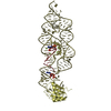
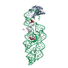
Mass: 4177.608 Da / Num. of mol.: 4 / Source method: obtained synthetically / Details: synthesized at Dharmacon / Source: (synth.) syntetic construct (others)
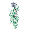
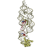
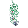
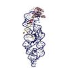
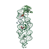
#3: RNA chain
glmSglucosamine-6-phosphateactivatedribozyme
Mass: 45608.859 Da / Num. of mol.: 4 / Source method: obtained synthetically / Details: in vitro transcribed from a DNA template / Source: (synth.) syntetic construct (others)
-
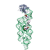
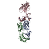
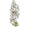
Protein / Sugars , 2 types, 8 molecules ABCD
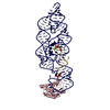
#1: Protein
U1smallnuclearribonucleoproteinA / U1 snRNP protein A / U1A protein / U1-A
Mass: 11340.315 Da / Num. of mol.: 4 / Fragment: RNA BINDING DOMAIN (UNP residues 1 to 98) / Mutation: Y31H,Q36R Source method: isolated from a genetically manipulated source Source: (gene. exp.) Homo sapiens (human) / Gene: SNRPA / Plasmid: pET11 / Production host: Escherichia coli (E. coli) / Strain (production host): BL21 / References: UniProt: P09012
In the structure databanks used in Yorodumi, some data are registered as the other names, "COVID-19 virus" and "2019-nCoV". Here are the details of the virus and the list of structure data.
Jan 31, 2019. EMDB accession codes are about to change! (news from PDBe EMDB page)
EMDB accession codes are about to change! (news from PDBe EMDB page)
The allocation of 4 digits for EMDB accession codes will soon come to an end. Whilst these codes will remain in use, new EMDB accession codes will include an additional digit and will expand incrementally as the available range of codes is exhausted. The current 4-digit format prefixed with “EMD-” (i.e. EMD-XXXX) will advance to a 5-digit format (i.e. EMD-XXXXX), and so on. It is currently estimated that the 4-digit codes will be depleted around Spring 2019, at which point the 5-digit format will come into force.
The EM Navigator/Yorodumi systems omit the EMD- prefix.
Related info.:Q: What is EMD? / ID/Accession-code notation in Yorodumi/EM Navigator
Yorodumi is a browser for structure data from EMDB, PDB, SASBDB, etc.
This page is also the successor to EM Navigator detail page, and also detail information page/front-end page for Omokage search.
The word "yorodu" (or yorozu) is an old Japanese word meaning "ten thousand". "mi" (miru) is to see.
Related info.:EMDB / PDB / SASBDB / Comparison of 3 databanks / Yorodumi Search / Aug 31, 2016. New EM Navigator & Yorodumi / Yorodumi Papers / Jmol/JSmol / Function and homology information / Changes in new EM Navigator and Yorodumi
 Movie
Movie Controller

 Yorodumi
 Open data
 Basic information
 Components
 Keywords
 Function and homology information
 Homo sapiens (human)
 X-RAY DIFFRACTION /
  Authors
 Citation
 Structure visualization
 Downloads & links
 Other downloads
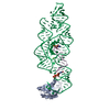


 PDBj
PDBj


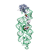
 Assembly

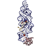
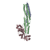
 Type: D-saccharide, alpha linking / Mass: 259.151 Da / Num. of mol.: 4

 Mass: 24.305 Da / Num. of mol.: 10 / Source method: obtained synthetically / Formula: Mg
 Sample preparation
 / Beamline: X29A / Wavelength: 1.1 Å
 Processing