Method to determine structure: MOLECULAR REPLACEMENT / Resolution: 1.76→34.03 Å / Cor.coef. Fo:Fc: 0.955 / Cor.coef. Fo:Fc free: 0.94 / SU B: 4.345 / SU ML: 0.07 / Cross valid method: THROUGHOUT / ESU R: 0.103 / ESU R Free: 0.101 / Stereochemistry target values: MAXIMUM LIKELIHOOD / Details: HYDROGENS HAVE BEEN USED IF PRESENT IN THE INPUT
Rfactor
Num. reflection
% reflection
Selection details
Rfree
0.2286
906
5 %
RANDOM
Rwork
0.20206
-
-
-
obs
0.20337
17163
98.05 %
-
Solvent computation
Ion probe radii: 0.8 Å / Shrinkage radii: 0.8 Å / VDW probe radii: 1.2 Å / Solvent model: BABINET MODEL WITH MASK
 Movie
Movie Controller


 Open data
 Basic information
 Components
 Keywords
 Function and homology information
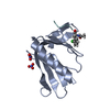
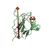
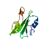
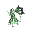
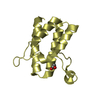
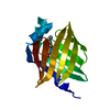
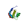
 Human cytomegalovirus
 X-RAY DIFFRACTION /
  Authors
 Citation
 Structure visualization
 Downloads & links
 Other downloads


 PDBj
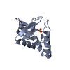
PDBj Assembly

 Mass: 18.015 Da / Num. of mol.: 74 / Source method: isolated from a natural source / Formula: H2O
 Sample preparation
 / Beamline: 14.1 / Wavelength: 0.91841 Å
 Processing