 Movie
Movie Controller


+ Open data

- Basic information
| Entry | Database: PDB / ID: 4or8 | ||||||
|---|---|---|---|---|---|---|---|
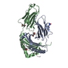
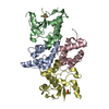
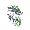
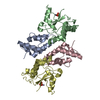
| Title | Crystal structure of Marburg virus VP24 | ||||||
 Components Components | Membrane-associated protein VP24 | ||||||
 Keywords Keywords | VIRAL PROTEIN / Marburg / VP24 / pyramidal fold / protein / Ebola / virus / viral | ||||||
| Function / homology |  Function and homology information Function and homology informationhost cell endomembrane system / viral process / viral nucleocapsid / structural constituent of virion / host cell plasma membrane / virion membrane / membrane Similarity search - Function | ||||||
| Biological species |  Marburg virus - Musoke Marburg virus - MusokeKenya 1980 | ||||||
| Method |  X-RAY DIFFRACTION / SYNCHROTRON / MOLECULAR REPLACEMENT / Resolution: 2.654 Å X-RAY DIFFRACTION / SYNCHROTRON / MOLECULAR REPLACEMENT / Resolution: 2.654 Å | ||||||
 Authors Authors | Zhang, A.P.P. / Bornholdt, Z. / Abelson, D. / Saphire, E.O. | ||||||
 Citation Citation | Journal: J.Virol. / Year: 2014 Title: Crystal Structure of Marburg Virus VP24. Authors: Zhang, A.P. / Bornholdt, Z.A. / Abelson, D.M. / Saphire, E.O. | ||||||
| History |
|
- Structure visualization
| Structure viewer | Molecule: MolmilJmol/JSmol |
|---|
- Downloads & links
-Download
| PDBx/mmCIF format | 4or8.cif.gz | 102.2 KB | Display | PDBx/mmCIF format |
|---|---|---|---|---|
| PDB format | pdb4or8.ent.gz | 78.6 KB | Display | PDB format |
| PDBx/mmJSON format | 4or8.json.gz | Tree view | PDBx/mmJSON format | |
| Others |  Other downloads Other downloads |
-Validation report
| Summary document | 4or8_validation.pdf.gz | 429.3 KB | Display | wwPDB validaton report |
|---|---|---|---|---|
| Full document | 4or8_full_validation.pdf.gz | 431.2 KB | Display | |
| Data in XML | 4or8_validation.xml.gz | 17.4 KB | Display | |
| Data in CIF | 4or8_validation.cif.gz | 23.2 KB | Display | |
| Arichive directory | https://data.pdbj.org/pub/pdb/validation_reports/or/4or8ftp://data.pdbj.org/pub/pdb/validation_reports/or/4or8 | HTTPS FTP |
-Related structure data
| Similar structure data |
|---|


-Links
 PDBj
PDBj
- Assembly
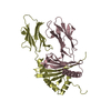
| Deposited unit | 
| ||||||||
|---|---|---|---|---|---|---|---|---|---|
| 1 |
| ||||||||
| Unit cell |
|
-Components
| #1: Protein | Mass: 30387.223 Da / Num. of mol.: 2 / Fragment: VP24 Source method: isolated from a genetically manipulated source Source: (gene. exp.) Marburg virus - Musoke, Kenya, 1980 / Strain: Musoke-80 / Gene: VP24 / Production host:  #2: Water | ChemComp-HOH / |  Mass: 18.015 Da / Num. of mol.: 47 / Source method: isolated from a natural source / Formula: H2O Mass: 18.015 Da / Num. of mol.: 47 / Source method: isolated from a natural source / Formula: H2O |
|---|
-Experimental details
-Experiment
| Experiment | Method: X-RAY DIFFRACTION / Number of used crystals: 1 |
|---|
- Sample preparation
| Crystal | Density Matthews: 1.99 Å3/Da / Density % sol: 38.07 % |
|---|---|
| Crystal grow | Temperature: 273 K / Method: liquid diffusion / pH: 6.5 Details: 0.1M ADA (Hampton Research), 0.1M lithium acetate, 20% glycerol, 2% (v/v) PEG 400 and 8% (v/v) PEG 4000 , pH 6.5, LIQUID DIFFUSION, temperature 273K |
-Data collection
| Diffraction | Mean temperature: 100 K |
|---|---|
| Diffraction source | Source: SYNCHROTRON / Site: ALS  / Beamline: 8.2.2 / Wavelength: 0.979 Å / Beamline: 8.2.2 / Wavelength: 0.979 Å |
| Detector | Type: ADSC QUANTUM 315 / Detector: CCD / Date: Oct 18, 2012 |
| Radiation | Monochromator: double crystal, Si(111) / Protocol: SINGLE WAVELENGTH / Monochromatic (M) / Laue (L): M / Scattering type: x-ray |
| Radiation wavelength | Wavelength: 0.979 Å / Relative weight: 1 |
| Reflection | Resolution: 2.654→40 Å / Num. all: 13515 / Num. obs: 13245 / % possible obs: 98 % / Observed criterion σ(F): 2 / Observed criterion σ(I): 4 / Redundancy: 2 % / Rmerge(I) obs: 0.06 / Net I/σ(I): 12.77 |
- Processing
| Software |
| ||||||||||||||||||||||||||||||||||||||||||
|---|---|---|---|---|---|---|---|---|---|---|---|---|---|---|---|---|---|---|---|---|---|---|---|---|---|---|---|---|---|---|---|---|---|---|---|---|---|---|---|---|---|---|---|
| Refinement | Method to determine structure: MOLECULAR REPLACEMENT / Resolution: 2.654→39.209 Å / SU ML: 0.35 / σ(F): 2.04 / Phase error: 26.21 / Stereochemistry target values: ML
| ||||||||||||||||||||||||||||||||||||||||||
| Solvent computation | Shrinkage radii: 0.9 Å / VDW probe radii: 1.11 Å / Solvent model: FLAT BULK SOLVENT MODEL | ||||||||||||||||||||||||||||||||||||||||||
| Refinement step | Cycle: LAST / Resolution: 2.654→39.209 Å
| ||||||||||||||||||||||||||||||||||||||||||
| Refine LS restraints |
| ||||||||||||||||||||||||||||||||||||||||||
| LS refinement shell |
|
