 Movie
Movie Controller

[English] 日本語
 Yorodumi
- PDB-4od2: Crystal structure of the Fab fragment of an anti-DR5 antibody bou... -
+ Open data

- Basic information
| Entry | Database: PDB / ID: 4od2 | ||||||
|---|---|---|---|---|---|---|---|
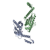
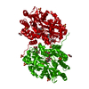
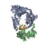
| Title | Crystal structure of the Fab fragment of an anti-DR5 antibody bound to DR5 | ||||||
 Components Components |
| ||||||
 Keywords Keywords | APOPTOSIS/IMMUNE SYSTEM / IgG / Fab fragment / CRD / TNFSF / APOPTOSIS-IMMUNE SYSTEM complex | ||||||
| Function / homology |  Function and homology information Function and homology informationTRAIL receptor activity / TRAIL binding / TRAIL signaling / TRAIL-activated apoptotic signaling pathway / Regulation by c-FLIP / CASP8 activity is inhibited / Dimerization of procaspase-8 / activation of NF-kappaB-inducing kinase activity / Caspase activation via Death Receptors in the presence of ligand / defense response to tumor cell ...TRAIL receptor activity / TRAIL binding / TRAIL signaling / TRAIL-activated apoptotic signaling pathway / Regulation by c-FLIP / CASP8 activity is inhibited / Dimerization of procaspase-8 / activation of NF-kappaB-inducing kinase activity / Caspase activation via Death Receptors in the presence of ligand / defense response to tumor cell / TP53 Regulates Transcription of Death Receptors and Ligands / RIPK1-mediated regulated necrosis / extrinsic apoptotic signaling pathway via death domain receptors / intrinsic apoptotic signaling pathway in response to endoplasmic reticulum stress / response to endoplasmic reticulum stress / Cell surface interactions at the vascular wall / cellular response to mechanical stimulus / signaling receptor activity / regulation of apoptotic process / positive regulation of canonical NF-kappaB signal transduction / cell surface receptor signaling pathway / positive regulation of apoptotic process / apoptotic process / cell surface / plasma membrane Similarity search - Function | ||||||
| Biological species |  Homo sapiens (human) Homo sapiens (human) | ||||||
| Method |  X-RAY DIFFRACTION / SYNCHROTRON / MOLECULAR REPLACEMENT / Resolution: 3.2 Å X-RAY DIFFRACTION / SYNCHROTRON / MOLECULAR REPLACEMENT / Resolution: 3.2 Å | ||||||
 Authors Authors | Hymowitz, S.G. / Compaan, D. | ||||||
 Citation Citation | Journal: Cell Death Differ. / Year: 2008 Title: Structural and functional analysis of the interaction between the agonistic monoclonal antibody Apomab and the proapoptotic receptor DR5. Authors: Adams, C. / Totpal, K. / Lawrence, D. / Marsters, S. / Pitti, R. / Yee, S. / Ross, S. / Deforge, L. / Koeppen, H. / Sagolla, M. / Compaan, D. / Lowman, H. / Hymowitz, S. / Ashkenazi, A. | ||||||
| History |
|
- Structure visualization
| Structure viewer | Molecule: MolmilJmol/JSmol |
|---|
- Downloads & links
-Download
| PDBx/mmCIF format | 4od2.cif.gz | 212.7 KB | Display | PDBx/mmCIF format |
|---|---|---|---|---|
| PDB format | pdb4od2.ent.gz | 173.2 KB | Display | PDB format |
| PDBx/mmJSON format | 4od2.json.gz | Tree view | PDBx/mmJSON format | |
| Others |  Other downloads Other downloads |
-Validation report
| Arichive directory | https://data.pdbj.org/pub/pdb/validation_reports/od/4od2ftp://data.pdbj.org/pub/pdb/validation_reports/od/4od2 | HTTPS FTP |
|---|
-Related structure data
| Related structure data | 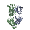 8fabS S: Starting model for refinement |
|---|---|
| Similar structure data |


-Links
 PDBj
PDBj


- Assembly
| Deposited unit | 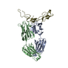
| ||||||||
|---|---|---|---|---|---|---|---|---|---|
| 1 |
| ||||||||
| Unit cell |
|
-Components
| #1: Antibody | Mass: 22418.611 Da / Num. of mol.: 1 Source method: isolated from a genetically manipulated source Source: (gene. exp.) Homo sapiens (human) / Production host:  |
|---|---|
| #2: Antibody | Mass: 24451.332 Da / Num. of mol.: 1 Source method: isolated from a genetically manipulated source Source: (gene. exp.) Homo sapiens (human) / Production host: |
| #3: Protein | Mass: 12488.968 Da / Num. of mol.: 1 / Fragment: UNP residues 73-183 Source method: isolated from a genetically manipulated source Source: (gene. exp.) Homo sapiens (human)Gene: Death receptor 5, DR5, KILLER, TNFRSF10B, TRAILR2, TRICK2, UNQ160/PRO186, ZTNFR9 Production host:   Spodoptera frugiperda (fall armyworm) / Strain (production host): Hi5 cells / References: UniProt: O14763 Spodoptera frugiperda (fall armyworm) / Strain (production host): Hi5 cells / References: UniProt: O14763 |
| Has protein modification | Y |
-Experimental details
-Experiment
| Experiment | Method: X-RAY DIFFRACTION |
|---|
- Sample preparation
| Crystal | Density Matthews: 2.74 Å3/Da / Density % sol: 55.08 % |
|---|---|
| Crystal grow | Temperature: 292 K / Method: vapor diffusion, hanging drop / pH: 8 Details: Protein (7mg/mL in 20 mM Tris-HCl, 150 mM NaCl) was mixed with equal volume of well solution (30% PEG 4K, 0.1 M Tris-HCl pH 8.5, 0.2M MgCl2), VAPOR DIFFUSION, HANGING DROP, temperature 292K |
-Data collection
| Diffraction | Mean temperature: 100 K |
|---|---|
| Diffraction source | Source: SYNCHROTRON / Site: ALS  / Beamline: 5.0.2 / Wavelength: 1 Å / Beamline: 5.0.2 / Wavelength: 1 Å |
| Detector | Type: ADSC QUANTUM 315 / Detector: CCD / Date: Apr 14, 2004 |
| Radiation | Monochromator: Si(111) / Protocol: SINGLE WAVELENGTH / Monochromatic (M) / Laue (L): M / Scattering type: x-ray |
| Radiation wavelength | Wavelength: 1 Å / Relative weight: 1 |
| Reflection | Resolution: 3.2→50 Å / Num. all: 10908 / Num. obs: 10792 / % possible obs: 98.9 % / Observed criterion σ(F): 0 / Observed criterion σ(I): -3 / Redundancy: 8.3 % / Rsym value: 0.087 / Net I/σ(I): 18.2 |
| Reflection shell | Resolution: 3.2→3.39 Å / Redundancy: 8.3 % / Mean I/σ(I) obs: 5.7 / Num. unique all: 1741 / Rsym value: 0.305 / % possible all: 96.8 |
- Processing
| Software |
| ||||||||||||||||||||||||||||||||||||||||||||||||||||||||||||||||||||||||||||||||||||||||||||||||||||||||||||||||||||||||||||||||||||||||||||||||||||||
|---|---|---|---|---|---|---|---|---|---|---|---|---|---|---|---|---|---|---|---|---|---|---|---|---|---|---|---|---|---|---|---|---|---|---|---|---|---|---|---|---|---|---|---|---|---|---|---|---|---|---|---|---|---|---|---|---|---|---|---|---|---|---|---|---|---|---|---|---|---|---|---|---|---|---|---|---|---|---|---|---|---|---|---|---|---|---|---|---|---|---|---|---|---|---|---|---|---|---|---|---|---|---|---|---|---|---|---|---|---|---|---|---|---|---|---|---|---|---|---|---|---|---|---|---|---|---|---|---|---|---|---|---|---|---|---|---|---|---|---|---|---|---|---|---|---|---|---|---|---|---|---|
| Refinement | Method to determine structure: MOLECULAR REPLACEMENT Starting model: PDB ENTRY 8FAB Resolution: 3.2→30 Å / Cor.coef. Fo:Fc: 0.899 / Cor.coef. Fo:Fc free: 0.829 / SU B: 73.538 / SU ML: 0.566 / Cross valid method: THROUGHOUT / ESU R Free: 0.654 / Stereochemistry target values: MAXIMUM LIKELIHOOD
| ||||||||||||||||||||||||||||||||||||||||||||||||||||||||||||||||||||||||||||||||||||||||||||||||||||||||||||||||||||||||||||||||||||||||||||||||||||||
| Solvent computation | Ion probe radii: 0.8 Å / Shrinkage radii: 0.8 Å / VDW probe radii: 1.4 Å / Solvent model: MASK | ||||||||||||||||||||||||||||||||||||||||||||||||||||||||||||||||||||||||||||||||||||||||||||||||||||||||||||||||||||||||||||||||||||||||||||||||||||||
| Displacement parameters | Biso mean: 111.762 Å2
| ||||||||||||||||||||||||||||||||||||||||||||||||||||||||||||||||||||||||||||||||||||||||||||||||||||||||||||||||||||||||||||||||||||||||||||||||||||||
| Refinement step | Cycle: LAST / Resolution: 3.2→30 Å
| ||||||||||||||||||||||||||||||||||||||||||||||||||||||||||||||||||||||||||||||||||||||||||||||||||||||||||||||||||||||||||||||||||||||||||||||||||||||
| Refine LS restraints |
| ||||||||||||||||||||||||||||||||||||||||||||||||||||||||||||||||||||||||||||||||||||||||||||||||||||||||||||||||||||||||||||||||||||||||||||||||||||||
| LS refinement shell | Resolution: 3.201→3.266 Å / Total num. of bins used: 25
| ||||||||||||||||||||||||||||||||||||||||||||||||||||||||||||||||||||||||||||||||||||||||||||||||||||||||||||||||||||||||||||||||||||||||||||||||||||||
| Refinement TLS params. | Method: refined / Refine-ID: X-RAY DIFFRACTION
| ||||||||||||||||||||||||||||||||||||||||||||||||||||||||||||||||||||||||||||||||||||||||||||||||||||||||||||||||||||||||||||||||||||||||||||||||||||||
| Refinement TLS group |
|
