| 登録情報 | データベース: PDB / ID: 4o8i
|
|---|
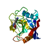
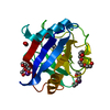
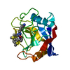
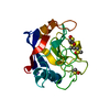
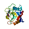
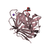
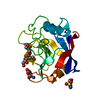
| タイトル | 1.45A resolution structure of PEG 400 Bound Cyclophilin D |
|---|
 要素 要素 | Peptidyl-prolyl cis-trans isomerase F, mitochondrial |
|---|
 キーワード キーワード | ISOMERASE |
|---|
| 機能・相同性 |  機能・相同性情報 機能・相同性情報
regulation of proton-transporting ATPase activity, rotational mechanism / negative regulation of oxidative phosphorylation uncoupler activity / mitochondrial outer membrane permeabilization involved in programmed cell death / regulation of mitochondrial membrane permeability involved in programmed necrotic cell death / skeletal muscle fiber differentiation / mitochondrial permeability transition pore complex / mitochondrial depolarization / cellular response to arsenic-containing substance / negative regulation of ATP-dependent activity / negative regulation of oxidative phosphorylation ...regulation of proton-transporting ATPase activity, rotational mechanism / negative regulation of oxidative phosphorylation uncoupler activity / mitochondrial outer membrane permeabilization involved in programmed cell death / regulation of mitochondrial membrane permeability involved in programmed necrotic cell death / skeletal muscle fiber differentiation / mitochondrial permeability transition pore complex / mitochondrial depolarization / cellular response to arsenic-containing substance / negative regulation of ATP-dependent activity / negative regulation of oxidative phosphorylation / regulation of mitochondrial membrane permeability / cyclosporin A binding / negative regulation of release of cytochrome c from mitochondria / apoptotic mitochondrial changes / necroptotic process / protein peptidyl-prolyl isomerization / negative regulation of intrinsic apoptotic signaling pathway / cellular response to calcium ion / peptidylprolyl isomerase / peptidyl-prolyl cis-trans isomerase activity / response to ischemia / cellular response to hydrogen peroxide / protein folding / mitochondrial inner membrane / mitochondrial matrix / negative regulation of apoptotic process / mitochondrion / membrane / cytoplasm類似検索 - 分子機能 Cyclophilin-like / Cyclophilin / Cyclophilin-type peptidyl-prolyl cis-trans isomerase, conserved site / Cyclophilin-type peptidyl-prolyl cis-trans isomerase signature. / Cyclophilin-type peptidyl-prolyl cis-trans isomerase domain profile. / Cyclophilin-type peptidyl-prolyl cis-trans isomerase domain / Cyclophilin type peptidyl-prolyl cis-trans isomerase/CLD / Cyclophilin-like domain superfamily / Beta Barrel / Mainly Beta類似検索 - ドメイン・相同性 Peptidyl-prolyl cis-trans isomerase F, mitochondrial類似検索 - 構成要素 |
|---|
| 生物種 |  Homo sapiens (ヒト) Homo sapiens (ヒト) |
|---|
| 手法 |  X線回折 / シンクロトロン / 分子置換 / 解像度: 1.45 Å X線回折 / シンクロトロン / 分子置換 / 解像度: 1.45 Å |
|---|
 データ登録者 データ登録者 | Lovell, S. / Valasani, K.R. / Battaile, K.P. / Wang, C. / Yan, S.S. |
|---|
 引用 引用 | ジャーナル: Acta Crystallogr F Struct Biol Commun / 年: 2014
タイトル: High-resolution crystal structures of two crystal forms of human cyclophilin D in complex with PEG 400 molecules.
著者: Valasani, K.R. / Carlson, E.A. / Battaile, K.P. / Bisson, A. / Wang, C. / Lovell, S. / ShiDu Yan, S. |
|---|
| 履歴 | | 登録 | 2013年12月27日 | 登録サイト: RCSB / 処理サイト: RCSB |
|---|
| 改定 1.0 | 2014年6月11日 | Provider: repository / タイプ: Initial release |
|---|
| 改定 1.1 | 2014年9月24日 | Group: Database references |
|---|
| 改定 1.2 | 2017年11月22日 | Group: Refinement description / カテゴリ: software / Item: _software.name |
|---|
| 改定 1.3 | 2023年9月20日 | Group: Data collection / Database references ...Data collection / Database references / Derived calculations / Refinement description
カテゴリ: chem_comp_atom / chem_comp_bond ...chem_comp_atom / chem_comp_bond / database_2 / pdbx_initial_refinement_model / struct_ref_seq_dif / struct_site
Item: _database_2.pdbx_DOI / _database_2.pdbx_database_accession ..._database_2.pdbx_DOI / _database_2.pdbx_database_accession / _struct_ref_seq_dif.details / _struct_site.pdbx_auth_asym_id / _struct_site.pdbx_auth_comp_id / _struct_site.pdbx_auth_seq_id |
|---|
|
|---|
 ムービー
ムービー コントローラー


 データを開く
 基本情報
基本情報 構造の表示
構造の表示 ダウンロードとリンク
 その他のダウンロード


 PDBj
PDBj

 集合体
集合体


 分子量: 238.278 Da / 分子数: 1 / 由来タイプ: 合成 / 式: C10H22O6 / コメント: 沈殿剤*YM
 分子量: 18.015 Da / 分子数: 133 / 由来タイプ: 天然 / 式: H2O
 試料調製
試料調製 / ビームライン: 17-ID / 波長: 1 Å
 解析
解析