 Movie
Movie Controller


+ Open data

- Basic information
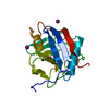
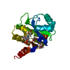
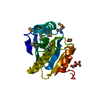
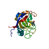
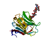
| Entry | Database: PDB / ID: 4b1m | |||||||||
|---|---|---|---|---|---|---|---|---|---|---|
| Title | CARBOHYDRATE BINDING MODULE CBM66 FROM BACILLUS SUBTILIS | |||||||||
 Components Components | LEVANASE | |||||||||
 Keywords Keywords | HYDROLASE / CBM66 | |||||||||
| Function / homology |  Function and homology information Function and homology informationfructan beta-fructosidase / fructan beta-fructosidase activity / sucrose catabolic process / sucrose alpha-glucosidase activity / extracellular region / cytoplasm Similarity search - Function | |||||||||
| Biological species |  | |||||||||
| Method |  X-RAY DIFFRACTION / SYNCHROTRON / MOLECULAR REPLACEMENT / Resolution: 1.1 Å X-RAY DIFFRACTION / SYNCHROTRON / MOLECULAR REPLACEMENT / Resolution: 1.1 Å | |||||||||
 Authors Authors | Cuskin, F. / Flint, J.E. / Morland, C. / Basle, A. / Henrissat, B. / Countinho, P.M. / Strazzulli, A. / Solzehinkin, A. / Davies, G.J. / Gilbert, H.J. / Gloster, T.M. | |||||||||
 Citation Citation | Journal: Proc.Natl.Acad.Sci.USA / Year: 2012 Title: How Nature Can Exploit Nonspecific Catalytic and Carbohydrate Binding Modules to Create Enzymatic Specificity Authors: Cuskin, F. / Flint, J.E. / Gloster, T.M. / Morland, C. / Basle, A. / Henrissat, B. / Coutinho, P.M. / Strazzulli, A. / Solovyova, A.S. / Davies, G.J. / Gilbert, H.J. | |||||||||
| History |
|
- Structure visualization
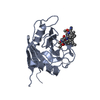
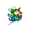
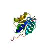
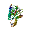
| Structure viewer | Molecule: MolmilJmol/JSmol |
|---|
- Downloads & links
-Download
| PDBx/mmCIF format | 4b1m.cif.gz | 227.9 KB | Display | PDBx/mmCIF format |
|---|---|---|---|---|
| PDB format | pdb4b1m.ent.gz | 180.2 KB | Display | PDB format |
| PDBx/mmJSON format | 4b1m.json.gz | Tree view | PDBx/mmJSON format | |
| Others |  Other downloads Other downloads |
-Validation report
| Arichive directory | https://data.pdbj.org/pub/pdb/validation_reports/b1/4b1mftp://data.pdbj.org/pub/pdb/validation_reports/b1/4b1m | HTTPS FTP |
|---|
-Related structure data
| Related structure data | 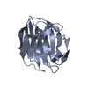 4azzSC 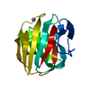 4b1lC S: Starting model for refinement C: citing same article ( |
|---|---|
| Similar structure data |

-Links
 PDBj
PDBj- Assembly
| Deposited unit | 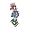
| ||||||||
|---|---|---|---|---|---|---|---|---|---|
| 1 | 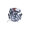
| ||||||||
| 2 | 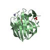
| ||||||||
| 3 | 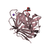
| ||||||||
| Unit cell |
|
-Components
| #1: Protein | Mass: 20643.711 Da / Num. of mol.: 3 / Fragment: CARBOHYDRATE BINDING MODULE, RESIDUES 515-677 Source method: isolated from a genetically manipulated source Details: CARBOHYDRATE BINDING MODULE WAS PREDICTED TO COMPRISE RESIDUES 515-677 OF THE FULL LENGTH PROTEIN. Source: (gene. exp.) #2: Polysaccharide | 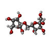  Source method: isolated from a genetically manipulated source Details: oligosaccharide / References: levanbiose #3: Chemical | ChemComp-NA /   Mass: 22.990 Da / Num. of mol.: 4 / Source method: obtained synthetically / Formula: Na Mass: 22.990 Da / Num. of mol.: 4 / Source method: obtained synthetically / Formula: Na#4: Chemical | ChemComp-SO4 / |   Mass: 96.063 Da / Num. of mol.: 1 / Source method: obtained synthetically / Formula: SO4 Mass: 96.063 Da / Num. of mol.: 1 / Source method: obtained synthetically / Formula: SO4#5: Water | ChemComp-HOH / |  Mass: 18.015 Da / Num. of mol.: 469 / Source method: isolated from a natural source / Formula: H2O Mass: 18.015 Da / Num. of mol.: 469 / Source method: isolated from a natural source / Formula: H2OSequence details | RESIDUES 515-677 OF THE FULL LENGTH PROTEIN WERE CLONED IN PET16B VECTOR. THE PROTEIN WAS DIGESTED ...RESIDUES 515-677 OF THE FULL LENGTH PROTEIN WERE CLONED IN PET16B VECTOR. THE PROTEIN WAS DIGESTED WITH FACTOR XA LEAVING ONLY HM RESIDUES IN N-TERMINAL OF THE TARGET SEQUENCE FOR CRYSTALLIS | |
|---|
-Experimental details
-Experiment
| Experiment | Method: X-RAY DIFFRACTION / Number of used crystals: 1 |
|---|
- Sample preparation
| Crystal | Density Matthews: 2.51 Å3/Da / Density % sol: 51 % / Description: NONE |
|---|---|
| Crystal grow | Details: 1.6-2.2 M AMMONIUM SULFATE, 1.4 - 2.4 M NACL, 5-6 MM LEVAN TRIOSE |
-Data collection
| Diffraction | Mean temperature: 100 K |
|---|---|
| Diffraction source | Source: SYNCHROTRON / Site: Diamond  / Beamline: I24 / Wavelength: 0.9784 / Beamline: I24 / Wavelength: 0.9784 |
| Detector | Type: DECTRIS PILATUS 6M / Detector: PIXEL / Date: Dec 18, 2011 |
| Radiation | Protocol: SINGLE WAVELENGTH / Monochromatic (M) / Laue (L): M / Scattering type: x-ray |
| Radiation wavelength | Wavelength: 0.9784 Å / Relative weight: 1 |
| Reflection | Resolution: 1.1→43.67 Å / Num. obs: 158032 / % possible obs: 91.2 % / Observed criterion σ(I): 2 / Redundancy: 3.3 % / Rmerge(I) obs: 0.05 / Net I/σ(I): 12.5 |
| Reflection shell | Resolution: 1.1→1.16 Å / Redundancy: 2.1 % / Rmerge(I) obs: 0.37 / Mean I/σ(I) obs: 2 / % possible all: 55.4 |
- Processing
| Software |
| ||||||||||||||||||||||||||||||||||||||||||||||||||||||||||||||||||||||||||||||||||||||||||||||||||||||||||||||||||||||||||||||||||||||||||||||||||||||||||||||||||||||||||||||||||||||
|---|---|---|---|---|---|---|---|---|---|---|---|---|---|---|---|---|---|---|---|---|---|---|---|---|---|---|---|---|---|---|---|---|---|---|---|---|---|---|---|---|---|---|---|---|---|---|---|---|---|---|---|---|---|---|---|---|---|---|---|---|---|---|---|---|---|---|---|---|---|---|---|---|---|---|---|---|---|---|---|---|---|---|---|---|---|---|---|---|---|---|---|---|---|---|---|---|---|---|---|---|---|---|---|---|---|---|---|---|---|---|---|---|---|---|---|---|---|---|---|---|---|---|---|---|---|---|---|---|---|---|---|---|---|---|---|---|---|---|---|---|---|---|---|---|---|---|---|---|---|---|---|---|---|---|---|---|---|---|---|---|---|---|---|---|---|---|---|---|---|---|---|---|---|---|---|---|---|---|---|---|---|---|---|
| Refinement | Method to determine structure: MOLECULAR REPLACEMENT Starting model: PDB ENTRY 4AZZ Resolution: 1.1→87.35 Å / Cor.coef. Fo:Fc: 0.974 / Cor.coef. Fo:Fc free: 0.967 / SU B: 1.269 / SU ML: 0.027 / Cross valid method: THROUGHOUT / ESU R: 0.036 / ESU R Free: 0.036 / Stereochemistry target values: MAXIMUM LIKELIHOOD Details: HYDROGENS HAVE BEEN ADDED IN THE RIDING POSITIONS. DISORDERED ATOMS WERE NOT MODELLED
| ||||||||||||||||||||||||||||||||||||||||||||||||||||||||||||||||||||||||||||||||||||||||||||||||||||||||||||||||||||||||||||||||||||||||||||||||||||||||||||||||||||||||||||||||||||||
| Solvent computation | Ion probe radii: 0.8 Å / Shrinkage radii: 0.8 Å / VDW probe radii: 1.2 Å / Solvent model: MASK | ||||||||||||||||||||||||||||||||||||||||||||||||||||||||||||||||||||||||||||||||||||||||||||||||||||||||||||||||||||||||||||||||||||||||||||||||||||||||||||||||||||||||||||||||||||||
| Displacement parameters | Biso mean: 9.975 Å2
| ||||||||||||||||||||||||||||||||||||||||||||||||||||||||||||||||||||||||||||||||||||||||||||||||||||||||||||||||||||||||||||||||||||||||||||||||||||||||||||||||||||||||||||||||||||||
| Refinement step | Cycle: LAST / Resolution: 1.1→87.35 Å
| ||||||||||||||||||||||||||||||||||||||||||||||||||||||||||||||||||||||||||||||||||||||||||||||||||||||||||||||||||||||||||||||||||||||||||||||||||||||||||||||||||||||||||||||||||||||
| Refine LS restraints |
|