 Movie
Movie Controller


+ Open data

- Basic information
| Entry | Database: PDB / ID: 4nsr | ||||||
|---|---|---|---|---|---|---|---|
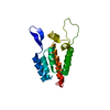
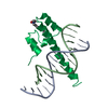
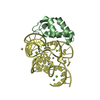
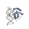
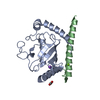
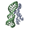
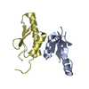
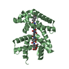
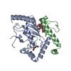
| Title | Crystal structure of the Immunity protein | ||||||
 Components Components | Immunity protein | ||||||
 Keywords Keywords | PROTEIN BINDING / Helix / peptidoglycan | ||||||
| Function / homology | Helicase, Ruva Protein; domain 3 - #1160 / : / : / Antitoxin TsiV3 / Helicase, Ruva Protein; domain 3 / Orthogonal Bundle / Mainly Alpha / identical protein binding / Antitoxin protein TsiV3 Function and homology information Function and homology information | ||||||
| Biological species |  Vibrio cholerae O1 biovar El Tor (bacteria) Vibrio cholerae O1 biovar El Tor (bacteria) | ||||||
| Method |  X-RAY DIFFRACTION / SYNCHROTRON / MOLECULAR REPLACEMENT / Resolution: 2.795 Å X-RAY DIFFRACTION / SYNCHROTRON / MOLECULAR REPLACEMENT / Resolution: 2.795 Å | ||||||
 Authors Authors | Dong, C. / Gao, Z.-Q. / Dong, Y.-H. | ||||||
 Citation Citation | Journal: Febs Lett. / Year: 2014 Title: Structural basis for recognition of the type VI spike protein VgrG3 by a cognate immunity protein. Authors: Zhang, J. / Zhang, H. / Gao, Z. / Hu, H. / Dong, C. / Dong, Y.H. | ||||||
| History |
|
- Structure visualization
| Structure viewer | Molecule: MolmilJmol/JSmol |
|---|
- Downloads & links
-Download
| PDBx/mmCIF format | 4nsr.cif.gz | 119.4 KB | Display | PDBx/mmCIF format |
|---|---|---|---|---|
| PDB format | pdb4nsr.ent.gz | 94.5 KB | Display | PDB format |
| PDBx/mmJSON format | 4nsr.json.gz | Tree view | PDBx/mmJSON format | |
| Others |  Other downloads Other downloads |
-Validation report
| Arichive directory | https://data.pdbj.org/pub/pdb/validation_reports/ns/4nsrftp://data.pdbj.org/pub/pdb/validation_reports/ns/4nsr | HTTPS FTP |
|---|
-Related structure data


-Links
 PDBj
PDBj- Assembly
| Deposited unit | 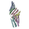
| ||||||||
|---|---|---|---|---|---|---|---|---|---|
| 1 | 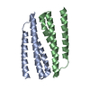
| ||||||||
| 2 | 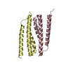
| ||||||||
| 3 | 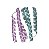
| ||||||||
| Unit cell |
| ||||||||
| Details | The asymmetric unit of a crystal structure contain three biological units. |
-Components
| #1: Protein | Mass: 13266.843 Da / Num. of mol.: 6 / Fragment: UNP residues 26-122 Source method: isolated from a genetically manipulated source Source: (gene. exp.) Vibrio cholerae O1 biovar El Tor (bacteria)Strain: N16961 / Gene: VC_A0124 / Production host: #2: Water | ChemComp-HOH / |  Mass: 18.015 Da / Num. of mol.: 38 / Source method: isolated from a natural source / Formula: H2O Mass: 18.015 Da / Num. of mol.: 38 / Source method: isolated from a natural source / Formula: H2O |
|---|
-Experimental details
-Experiment
| Experiment | Method: X-RAY DIFFRACTION / Number of used crystals: 1 |
|---|
- Sample preparation
| Crystal | Density Matthews: 1.94 Å3/Da / Density % sol: 36.69 % |
|---|---|
| Crystal grow | Temperature: 277 K / Method: vapor diffusion, sitting drop / pH: 7 Details: 0.2M Ammonium citrate tribasic, 20% w/v Polyethylene glycol monomethyl ether 2000, 0.1M Imidazole pH7.0, VAPOR DIFFUSION, SITTING DROP, temperature 277K |
-Data collection
| Diffraction | Mean temperature: 100 K |
|---|---|
| Diffraction source | Source: SYNCHROTRON / Site: BSRF  / Beamline: 3W1A / Wavelength: 1 Å / Beamline: 3W1A / Wavelength: 1 Å |
| Detector | Type: MAR CCD 165 mm / Detector: CCD / Date: Oct 15, 2013 |
| Radiation | Monochromator: double crystal / Protocol: SINGLE WAVELENGTH / Monochromatic (M) / Laue (L): M / Scattering type: x-ray |
| Radiation wavelength | Wavelength: 1 Å / Relative weight: 1 |
| Reflection | Resolution: 2.795→50 Å / Num. obs: 15700 / Redundancy: 5.9 % / Rmerge(I) obs: 0.077 / Net I/σ(I): 25 |
| Reflection shell | Resolution: 2.8→2.85 Å / Redundancy: 5.7 % / Rmerge(I) obs: 0.401 / Mean I/σ(I) obs: 3 / Num. unique all: 770 / % possible all: 99.4 |
- Processing
| Software |
| |||||||||||||||||||||||||||||||||||||||||||||||||
|---|---|---|---|---|---|---|---|---|---|---|---|---|---|---|---|---|---|---|---|---|---|---|---|---|---|---|---|---|---|---|---|---|---|---|---|---|---|---|---|---|---|---|---|---|---|---|---|---|---|---|
| Refinement | Method to determine structure: MOLECULAR REPLACEMENT / Resolution: 2.795→44.115 Å / SU ML: 0.54 / σ(F): 1.36 / Phase error: 30.81 / Stereochemistry target values: ML
| |||||||||||||||||||||||||||||||||||||||||||||||||
| Solvent computation | Shrinkage radii: 0.98 Å / VDW probe radii: 1.2 Å / Solvent model: FLAT BULK SOLVENT MODEL / Bsol: 42.282 Å2 / ksol: 0.328 e/Å3 | |||||||||||||||||||||||||||||||||||||||||||||||||
| Displacement parameters |
| |||||||||||||||||||||||||||||||||||||||||||||||||
| Refinement step | Cycle: LAST / Resolution: 2.795→44.115 Å
| |||||||||||||||||||||||||||||||||||||||||||||||||
| Refine LS restraints |
| |||||||||||||||||||||||||||||||||||||||||||||||||
| LS refinement shell |
|
