 Movie
Movie Controller


+ Open data

- Basic information
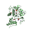
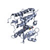
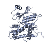
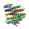
| Entry | Database: PDB / ID: 6op8 | |||||||||||||||
|---|---|---|---|---|---|---|---|---|---|---|---|---|---|---|---|---|
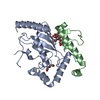
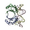
| Title | S. pombe Ubc7/U7BR complex | |||||||||||||||
 Components Components |
| |||||||||||||||
 Keywords Keywords | LIGASE/PROTEIN BINDING / UBL CONJUGATION PATHWAY / ENDOPLASMIC RETICULUM-ASSOCIATED DEGRADATION / LIGASE / LIGASE-PROTEIN BINDING complex | |||||||||||||||
| Function / homology |  Function and homology information Function and homology informationinner nuclear membrane-associated protein degradation pathway / Hrd1p ubiquitin ligase complex / : / Antigen processing: Ubiquitination & Proteasome degradation / ubiquitin-protein transferase activator activity / E2 ubiquitin-conjugating enzyme / ubiquitin conjugating enzyme activity / ERAD pathway / ubiquitin binding / protein ubiquitination ...inner nuclear membrane-associated protein degradation pathway / Hrd1p ubiquitin ligase complex / : / Antigen processing: Ubiquitination & Proteasome degradation / ubiquitin-protein transferase activator activity / E2 ubiquitin-conjugating enzyme / ubiquitin conjugating enzyme activity / ERAD pathway / ubiquitin binding / protein ubiquitination / endoplasmic reticulum / mitochondrion / ATP binding / nucleus / cytosol Similarity search - Function | |||||||||||||||
| Biological species |  | |||||||||||||||
| Method |  X-RAY DIFFRACTION / SYNCHROTRON / MOLECULAR REPLACEMENT / Resolution: 1.703 Å X-RAY DIFFRACTION / SYNCHROTRON / MOLECULAR REPLACEMENT / Resolution: 1.703 Å | |||||||||||||||
 Authors Authors | Hann, Z.S. / Lima, C.D. | |||||||||||||||
| Funding support |  United States, 4items United States, 4items
| |||||||||||||||
 Citation Citation | Journal: Acta Crystallogr.,Sect.F / Year: 2019 Title: Crystal structure of the Schizosaccharomyces pombe U7BR E2-binding region in complex with Ubc7. Authors: Hann, Z.S. / Metzger, M.B. / Weissman, A.M. / Lima, C.D. | |||||||||||||||
| History |
|
- Structure visualization
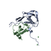
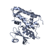
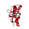
| Structure viewer | Molecule: MolmilJmol/JSmol |
|---|
- Downloads & links
-Download
| PDBx/mmCIF format | 6op8.cif.gz | 65.7 KB | Display | PDBx/mmCIF format |
|---|---|---|---|---|
| PDB format | pdb6op8.ent.gz | 45.6 KB | Display | PDB format |
| PDBx/mmJSON format | 6op8.json.gz | Tree view | PDBx/mmJSON format | |
| Others |  Other downloads Other downloads |
-Validation report
| Arichive directory | https://data.pdbj.org/pub/pdb/validation_reports/op/6op8ftp://data.pdbj.org/pub/pdb/validation_reports/op/6op8 | HTTPS FTP |
|---|
-Related structure data
| Related structure data |  4jquS S: Starting model for refinement |
|---|---|
| Similar structure data |
-Links
 PDBj
PDBj

- Assembly
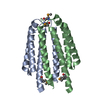
| Deposited unit | 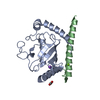
| ||||||||
|---|---|---|---|---|---|---|---|---|---|
| 1 |
| ||||||||
| Unit cell |
|
-Components
-Protein , 2 types, 2 molecules AB
| #1: Protein | Mass: 19206.811 Da / Num. of mol.: 1 Source method: isolated from a genetically manipulated source Source: (gene. exp.) Strain: 972 / ATCC 24843 / Gene: ubc7, ubcp3, SPBP16F5.04 / Plasmid: pET29b / Production host:  References: UniProt: O00102, E2 ubiquitin-conjugating enzyme |
|---|---|
| #2: Protein | Mass: 7814.836 Da / Num. of mol.: 1 / Fragment: residues 152-215 Source method: isolated from a genetically manipulated source Source: (gene. exp.) Strain: 972 / ATCC 24843 / Gene: SPCC4G3.13c / Plasmid: pET28b / Production host: |
-Non-polymers , 4 types, 217 molecules 


| #3: Chemical | ChemComp-MG /  Mass: 24.305 Da / Num. of mol.: 1 / Source method: obtained synthetically / Formula: Mg Mass: 24.305 Da / Num. of mol.: 1 / Source method: obtained synthetically / Formula: Mg |
|---|---|
| #4: Chemical | ChemComp-EDO /  Mass: 62.068 Da / Num. of mol.: 1 / Source method: obtained synthetically / Formula: C2H6O2 Mass: 62.068 Da / Num. of mol.: 1 / Source method: obtained synthetically / Formula: C2H6O2 |
| #5: Chemical | ChemComp-NA /  Mass: 22.990 Da / Num. of mol.: 1 / Source method: obtained synthetically / Formula: Na Mass: 22.990 Da / Num. of mol.: 1 / Source method: obtained synthetically / Formula: Na |
| #6: Water | ChemComp-HOH / Mass: 18.015 Da / Num. of mol.: 214 / Source method: isolated from a natural source / Formula: H2O |
-Experimental details
-Experiment
| Experiment | Method: X-RAY DIFFRACTION / Number of used crystals: 1 |
|---|
- Sample preparation
| Crystal | Density Matthews: 1.85 Å3/Da / Density % sol: 33.39 % |
|---|---|
| Crystal grow | Temperature: 291 K / Method: vapor diffusion, hanging drop / pH: 8 / Details: 100 mM TRIS, pH 8.0, 200 mM MgCl2, 30% PEG4000 |
-Data collection
| Diffraction | Mean temperature: 100 K / Serial crystal experiment: N |
|---|---|
| Diffraction source | Source: SYNCHROTRON / Site: APS / Beamline: 24-ID-C / Wavelength: 0.9791 Å |
| Detector | Type: DECTRIS PILATUS 6M-F / Detector: PIXEL / Date: Nov 8, 2015 |
| Radiation | Protocol: SINGLE WAVELENGTH / Monochromatic (M) / Laue (L): M / Scattering type: x-ray |
| Radiation wavelength | Wavelength: 0.9791 Å / Relative weight: 1 |
| Reflection | Resolution: 1.703→47.2787 Å / Num. obs: 22673 / % possible obs: 99.39 % / Redundancy: 5.7 % / Biso Wilson estimate: 14.49 Å2 / CC1/2: 0.999 / Rmerge(I) obs: 0.0562 / Rpim(I) all: 0.02523 / Rrim(I) all: 0.06177 / Net I/σ(I): 25.94 |
| Reflection shell | Resolution: 1.703→1.764 Å / Redundancy: 5.7 % / Rmerge(I) obs: 0.2232 / Mean I/σ(I) obs: 9.05 / Num. unique obs: 2195 / CC1/2: 0.97 / Rpim(I) all: 0.1023 / Rrim(I) all: 0.2461 / % possible all: 98.12 |
- Processing
| Software |
| |||||||||||||||||||||||||||||||||||||||||||||||||||||||||||||||
|---|---|---|---|---|---|---|---|---|---|---|---|---|---|---|---|---|---|---|---|---|---|---|---|---|---|---|---|---|---|---|---|---|---|---|---|---|---|---|---|---|---|---|---|---|---|---|---|---|---|---|---|---|---|---|---|---|---|---|---|---|---|---|---|---|
| Refinement | Method to determine structure: MOLECULAR REPLACEMENT Starting model: 4JQU Resolution: 1.703→47.26 Å / SU ML: 0.17 / Cross valid method: THROUGHOUT / σ(F): 1.38 / Phase error: 18.92
| |||||||||||||||||||||||||||||||||||||||||||||||||||||||||||||||
| Solvent computation | Shrinkage radii: 0.9 Å / VDW probe radii: 1.11 Å | |||||||||||||||||||||||||||||||||||||||||||||||||||||||||||||||
| Displacement parameters | Biso max: 106.47 Å2 / Biso mean: 20.4469 Å2 / Biso min: 3.7 Å2 | |||||||||||||||||||||||||||||||||||||||||||||||||||||||||||||||
| Refinement step | Cycle: final / Resolution: 1.703→47.26 Å
| |||||||||||||||||||||||||||||||||||||||||||||||||||||||||||||||
| LS refinement shell | Refine-ID: X-RAY DIFFRACTION / Rfactor Rfree error: 0 / Total num. of bins used: 8
|
