 Movie
Movie Controller

[English] 日本語
 Yorodumi
- PDB-4nm7: Crystal structure of GSK-3/Axin complex bound to phosphorylated W... -
+ Open data

- Basic information
| Entry | Database: PDB / ID: 4nm7 | ||||||
|---|---|---|---|---|---|---|---|
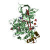
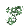
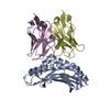
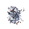
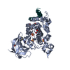
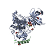
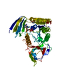
| Title | Crystal structure of GSK-3/Axin complex bound to phosphorylated Wnt receptor LRP6 e-motif | ||||||
 Components Components |
| ||||||
 Keywords Keywords | TRANSFERASE/PEPTIDE / Wnt / LRP6 / Auto-inhibited / GSK-3 / primed substrate / Kinase / Axin / phosphorylated Wnt receptor LRP6 e-motif / TRANSFERASE-PEPTIDE complex | ||||||
| Function / homology |  Function and homology information Function and homology informationbeta-catenin destruction complex assembly / cellular response to oxygen-containing compound / positive regulation of establishment of protein localization / Wnt-Frizzled-LRP5/6 complex / armadillo repeat domain binding / Negative regulation of TCF-dependent signaling by WNT ligand antagonists / Signaling by RNF43 mutants / neural crest formation / kinase inhibitor activity / head development ...beta-catenin destruction complex assembly / cellular response to oxygen-containing compound / positive regulation of establishment of protein localization / Wnt-Frizzled-LRP5/6 complex / armadillo repeat domain binding / Negative regulation of TCF-dependent signaling by WNT ligand antagonists / Signaling by RNF43 mutants / neural crest formation / kinase inhibitor activity / head development / cell development / negative regulation of glycogen (starch) synthase activity / axial mesoderm formation / neuron projection organization / regulation of microtubule anchoring at centrosome / negative regulation of mesenchymal stem cell differentiation / negative regulation of type B pancreatic cell development / dorsal/ventral axis specification / Wnt receptor activity / superior temporal gyrus development / positive regulation of protein localization to cilium / negative regulation of glycogen biosynthetic process / negative regulation of TORC2 signaling / low-density lipoprotein particle receptor activity / beta-arrestin-dependent dopamine receptor signaling pathway / toxin transmembrane transporter activity / Wnt-protein binding / negative regulation of dopaminergic neuron differentiation / positive regulation of protein localization to centrosome / maintenance of cell polarity / cellular response to cholesterol / positive regulation of cilium assembly / post-anal tail morphogenesis / CRMPs in Sema3A signaling / tau-protein kinase / heart valve development / beta-catenin destruction complex / dopaminergic neuron differentiation / midbrain dopaminergic neuron differentiation / APC truncation mutants have impaired AXIN binding / AXIN missense mutants destabilize the destruction complex / Truncations of AMER1 destabilize the destruction complex / positive regulation of mitochondrial outer membrane permeabilization involved in apoptotic signaling pathway / regulation of protein export from nucleus / cellular response to interleukin-3 / Maturation of nucleoprotein / Beta-catenin phosphorylation cascade / Signaling by GSK3beta mutants / CTNNB1 S33 mutants aren't phosphorylated / CTNNB1 S37 mutants aren't phosphorylated / CTNNB1 S45 mutants aren't phosphorylated / CTNNB1 T41 mutants aren't phosphorylated / frizzled binding / I-SMAD binding / regulation of long-term synaptic potentiation / Wnt signalosome / negative regulation of TOR signaling / regulation of microtubule-based process / AKT phosphorylates targets in the cytosol / negative regulation of protein metabolic process / Disassembly of the destruction complex and recruitment of AXIN to the membrane / positive regulation of protein binding / epigenetic programming in the zygotic pronuclei / regulation of axon extension / negative regulation of calcineurin-NFAT signaling cascade / positive regulation of ubiquitin-dependent protein catabolic process / Maturation of nucleoprotein / nucleocytoplasmic transport / negative regulation of protein localization to nucleus / RUNX1 regulates transcription of genes involved in WNT signaling / RUNX1 regulates estrogen receptor mediated transcription / negative regulation of epithelial to mesenchymal transition / neural crest cell differentiation / tau-protein kinase activity / positive regulation of cell-matrix adhesion / glycogen metabolic process / ER overload response / regulation of axonogenesis / negative regulation of fat cell differentiation / regulation of dendrite morphogenesis / regulation of neuron projection development / Constitutive Signaling by AKT1 E17K in Cancer / positive regulation of transforming growth factor beta receptor signaling pathway / protein kinase A catalytic subunit binding / SMAD binding / establishment of cell polarity / negative regulation of smooth muscle cell apoptotic process / R-SMAD binding / negative regulation of transcription elongation by RNA polymerase II / dynactin binding / epithelial to mesenchymal transition / lateral plasma membrane / Regulation of HSF1-mediated heat shock response / canonical Wnt signaling pathway / negative regulation of signal transduction / protein serine/threonine kinase inhibitor activity / negative regulation of osteoblast differentiation / NF-kappaB binding / negative regulation of extrinsic apoptotic signaling pathway via death domain receptors / negative regulation of protein-containing complex assembly Similarity search - Function | ||||||
| Biological species |  Homo sapiens (human) Homo sapiens (human) | ||||||
| Method |  X-RAY DIFFRACTION / SYNCHROTRON / MOLECULAR REPLACEMENT / molecular replacement / Resolution: 2.3 Å X-RAY DIFFRACTION / SYNCHROTRON / MOLECULAR REPLACEMENT / molecular replacement / Resolution: 2.3 Å | ||||||
 Authors Authors | Stamos, J.L. / Chu, M.L.-H. / Enos, M.D. / Shah, N. / Weis, W.I. | ||||||
 Citation Citation | Journal: Elife / Year: 2014 Title: Structural basis of GSK-3 inhibition by N-terminal phosphorylation and by the Wnt receptor LRP6. Authors: Stamos, J.L. / Chu, M.L. / Enos, M.D. / Shah, N. / Weis, W.I. | ||||||
| History |
|
- Structure visualization
| Structure viewer | Molecule: MolmilJmol/JSmol |
|---|
- Downloads & links
-Download
| PDBx/mmCIF format | 4nm7.cif.gz | 177.6 KB | Display | PDBx/mmCIF format |
|---|---|---|---|---|
| PDB format | pdb4nm7.ent.gz | 139.5 KB | Display | PDB format |
| PDBx/mmJSON format | 4nm7.json.gz | Tree view | PDBx/mmJSON format | |
| Others |  Other downloads Other downloads |
-Validation report
| Arichive directory | https://data.pdbj.org/pub/pdb/validation_reports/nm/4nm7ftp://data.pdbj.org/pub/pdb/validation_reports/nm/4nm7 | HTTPS FTP |
|---|
-Related structure data
| Related structure data |  4nm0C  4nm3C  4nm5C  4nu1C  1o9uS C: citing same article ( S: Starting model for refinement |
|---|---|
| Similar structure data |


-Links
 PDBj
PDBj


- Assembly
| Deposited unit | 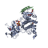
| ||||||||
|---|---|---|---|---|---|---|---|---|---|
| 1 |
| ||||||||
| Unit cell |
| ||||||||
| Components on special symmetry positions |
|
-Components
-Protein , 1 types, 1 molecules A
| #1: Protein | Mass: 42857.160 Da / Num. of mol.: 1 / Fragment: Residues 13-383 Source method: isolated from a genetically manipulated source Source: (gene. exp.) Homo sapiens (human) / Gene: GSK3B, hCG_1818062 / Plasmid: pET29b(+) / Production host:  References: UniProt: Q6FI27, UniProt: P49841*PLUS, tau-protein kinase |
|---|
-Protein/peptide , 2 types, 2 molecules BC
| #2: Protein/peptide | Mass: 2738.144 Da / Num. of mol.: 1 / Fragment: Residues 383-402 Source method: isolated from a genetically manipulated source Source: (gene. exp.) Homo sapiens (human) / Gene: AXIN1, AXIN / Plasmid: Modified pGEX-KG / Production host: |
|---|---|
| #3: Protein/peptide | Mass: 1006.089 Da / Num. of mol.: 1 / Fragment: Residues 1603-1610 Source method: isolated from a genetically manipulated source Source: (gene. exp.) Homo sapiens (human) / Plasmid: pET29b(+) / Production host: |
-Non-polymers , 6 types, 114 molecules 


| #4: Chemical | ChemComp-ADP /  Mass: 427.201 Da / Num. of mol.: 1 / Source method: obtained synthetically / Formula: C10H15N5O10P2 / Comment: ADP, energy-carrying molecule*YM Mass: 427.201 Da / Num. of mol.: 1 / Source method: obtained synthetically / Formula: C10H15N5O10P2 / Comment: ADP, energy-carrying molecule*YM | ||||||||
|---|---|---|---|---|---|---|---|---|---|
| #5: Chemical |  Mass: 92.094 Da / Num. of mol.: 3 / Source method: obtained synthetically / Formula: C3H8O3 Mass: 92.094 Da / Num. of mol.: 3 / Source method: obtained synthetically / Formula: C3H8O3#6: Chemical |  Mass: 24.305 Da / Num. of mol.: 2 / Source method: obtained synthetically / Formula: Mg Mass: 24.305 Da / Num. of mol.: 2 / Source method: obtained synthetically / Formula: Mg#7: Chemical | ChemComp-CL / |  Mass: 35.453 Da / Num. of mol.: 1 / Source method: obtained synthetically / Formula: Cl Mass: 35.453 Da / Num. of mol.: 1 / Source method: obtained synthetically / Formula: Cl#8: Chemical | ChemComp-DTT / |  Mass: 154.251 Da / Num. of mol.: 1 / Source method: obtained synthetically / Formula: C4H10O2S2 Mass: 154.251 Da / Num. of mol.: 1 / Source method: obtained synthetically / Formula: C4H10O2S2#9: Water | ChemComp-HOH / | Mass: 18.015 Da / Num. of mol.: 106 / Source method: isolated from a natural source / Formula: H2O |
-Details
| Has protein modification | Y |
|---|
-Experimental details
-Experiment
| Experiment | Method: X-RAY DIFFRACTION / Number of used crystals: 1 |
|---|
- Sample preparation
| Crystal | Density Matthews: 2.9 Å3/Da / Density % sol: 57.57 % |
|---|---|
| Crystal grow | Temperature: 277 K / pH: 7.5 Details: 10% PEG 35,000, 20mM Tris 7.5, 300mM NaCl, 5% glycerol, 10mM MgCl2, 200uM ATP, and 5mM DTT, MICRODIALYSIS, temperature 277K |
-Data collection
| Diffraction | Mean temperature: 100 K |
|---|---|
| Diffraction source | Source: SYNCHROTRON / Site: SSRL  / Beamline: BL12-2 / Wavelength: 1 / Beamline: BL12-2 / Wavelength: 1 |
| Detector | Type: PSI PILATUS 6M / Detector: PIXEL / Date: Mar 31, 2013 |
| Radiation | Monochromator: SI(III) / Protocol: SINGLE WAVELENGTH / Monochromatic (M) / Laue (L): M / Scattering type: x-ray |
| Radiation wavelength | Wavelength: 1 Å / Relative weight: 1 |
| Reflection | Resolution: 2.3→39.352 Å / Num. obs: 25932 / % possible obs: 100 % / Redundancy: 18.7 % / Biso Wilson estimate: 60.05 Å2 / Rsym value: 0.136 / Net I/σ(I): 17.8 |
| Reflection shell | Resolution: 2.3→2.38 Å / Redundancy: 18.2 % / Mean I/σ(I) obs: 0.9 / Rsym value: 4.307 / % possible all: 100 |
-Phasing
| Phasing | Method: molecular replacement |
|---|
- Processing
| Software |
| |||||||||||||||||||||||||||||||||||||||||||||||||||||||||||||||||||||||||||||||||||||||||||||||||||||||||||||||||||||||||||||||||||||||||||||||||||||||||||||||||||||||||||||||
|---|---|---|---|---|---|---|---|---|---|---|---|---|---|---|---|---|---|---|---|---|---|---|---|---|---|---|---|---|---|---|---|---|---|---|---|---|---|---|---|---|---|---|---|---|---|---|---|---|---|---|---|---|---|---|---|---|---|---|---|---|---|---|---|---|---|---|---|---|---|---|---|---|---|---|---|---|---|---|---|---|---|---|---|---|---|---|---|---|---|---|---|---|---|---|---|---|---|---|---|---|---|---|---|---|---|---|---|---|---|---|---|---|---|---|---|---|---|---|---|---|---|---|---|---|---|---|---|---|---|---|---|---|---|---|---|---|---|---|---|---|---|---|---|---|---|---|---|---|---|---|---|---|---|---|---|---|---|---|---|---|---|---|---|---|---|---|---|---|---|---|---|---|---|---|---|---|
| Refinement | Method to determine structure: MOLECULAR REPLACEMENT Starting model: PDB ENTRY 1O9U Resolution: 2.3→39.35 Å / Occupancy max: 1 / Occupancy min: 0.46 / SU ML: 0.37 / σ(F): 1.91 / Phase error: 24.14 / Stereochemistry target values: ML
| |||||||||||||||||||||||||||||||||||||||||||||||||||||||||||||||||||||||||||||||||||||||||||||||||||||||||||||||||||||||||||||||||||||||||||||||||||||||||||||||||||||||||||||||
| Solvent computation | Shrinkage radii: 0.9 Å / VDW probe radii: 1.11 Å / Solvent model: FLAT BULK SOLVENT MODEL | |||||||||||||||||||||||||||||||||||||||||||||||||||||||||||||||||||||||||||||||||||||||||||||||||||||||||||||||||||||||||||||||||||||||||||||||||||||||||||||||||||||||||||||||
| Displacement parameters | Biso mean: 70.4 Å2 | |||||||||||||||||||||||||||||||||||||||||||||||||||||||||||||||||||||||||||||||||||||||||||||||||||||||||||||||||||||||||||||||||||||||||||||||||||||||||||||||||||||||||||||||
| Refinement step | Cycle: LAST / Resolution: 2.3→39.35 Å
| |||||||||||||||||||||||||||||||||||||||||||||||||||||||||||||||||||||||||||||||||||||||||||||||||||||||||||||||||||||||||||||||||||||||||||||||||||||||||||||||||||||||||||||||
| Refine LS restraints |
| |||||||||||||||||||||||||||||||||||||||||||||||||||||||||||||||||||||||||||||||||||||||||||||||||||||||||||||||||||||||||||||||||||||||||||||||||||||||||||||||||||||||||||||||
| LS refinement shell |
| |||||||||||||||||||||||||||||||||||||||||||||||||||||||||||||||||||||||||||||||||||||||||||||||||||||||||||||||||||||||||||||||||||||||||||||||||||||||||||||||||||||||||||||||
| Refinement TLS params. | Method: refined / Refine-ID: X-RAY DIFFRACTION
| |||||||||||||||||||||||||||||||||||||||||||||||||||||||||||||||||||||||||||||||||||||||||||||||||||||||||||||||||||||||||||||||||||||||||||||||||||||||||||||||||||||||||||||||
| Refinement TLS group |
|
