 Movie
Movie Controller

[English] 日本語
 Yorodumi
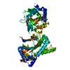
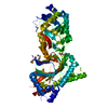
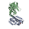
- PDB-4nhl: Crystal structure of Tpa1p from Saccharomyces cerevisiae, termina... -
+ Open data

- Basic information
| Entry | Database: PDB / ID: 4nhl | ||||||
|---|---|---|---|---|---|---|---|
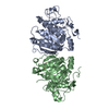
| Title | Crystal structure of Tpa1p from Saccharomyces cerevisiae, termination and polyadenylation protein 1, in complex with N-oxalylglycine (NOG) | ||||||
 Components Components | PKHD-type hydroxylase TPA1 | ||||||
 Keywords Keywords | Oxidoreductase/Oxidoreductase inhibitor / 2-oxoglutarate oxygenase / oxygen sensing / protein synthesis regulation / double-stranded beta helix / jellyroll fold / prolyl hydroxylase / translation / ribosome / Oxidoreductase-Oxidoreductase inhibitor complex | ||||||
| Function / homology |  Function and homology information Function and homology informationpeptidyl-proline di-hydroxylation / peptidyl-proline dioxygenase activity / regulation of translational termination / poly(A) binding / Oxidoreductases; Acting on paired donors, with incorporation or reduction of molecular oxygen; With 2-oxoglutarate as one donor, and incorporation of one atom of oxygen into each donor / L-ascorbic acid binding / Protein hydroxylation / nuclear-transcribed mRNA catabolic process, deadenylation-dependent decay / translational termination / regulation of translational fidelity ...peptidyl-proline di-hydroxylation / peptidyl-proline dioxygenase activity / regulation of translational termination / poly(A) binding / Oxidoreductases; Acting on paired donors, with incorporation or reduction of molecular oxygen; With 2-oxoglutarate as one donor, and incorporation of one atom of oxygen into each donor / L-ascorbic acid binding / Protein hydroxylation / nuclear-transcribed mRNA catabolic process, deadenylation-dependent decay / translational termination / regulation of translational fidelity / ferrous iron binding / nucleus / cytoplasm Similarity search - Function | ||||||
| Biological species |  | ||||||
| Method |  X-RAY DIFFRACTION / MOLECULAR REPLACEMENT / molecular replacement / Resolution: 2.84 Å X-RAY DIFFRACTION / MOLECULAR REPLACEMENT / molecular replacement / Resolution: 2.84 Å | ||||||
 Authors Authors | Scotti, J.S. / McDonough, M.A. / Schofield, C.J. | ||||||
 Citation Citation | Journal: Structure / Year: 2015 Title: Structure of the Ribosomal Oxygenase OGFOD1 Provides Insights into the Regio- and Stereoselectivity of Prolyl Hydroxylases. Authors: Horita, S. / Scotti, J.S. / Thinnes, C. / Mottaghi-Taromsari, Y.S. / Thalhammer, A. / Ge, W. / Aik, W. / Loenarz, C. / Schofield, C.J. / McDonough, M.A. | ||||||
| History |
|
- Structure visualization
| Structure viewer | Molecule: MolmilJmol/JSmol |
|---|
- Downloads & links
-Download
| PDBx/mmCIF format | 4nhl.cif.gz | 232.9 KB | Display | PDBx/mmCIF format |
|---|---|---|---|---|
| PDB format | pdb4nhl.ent.gz | 185.8 KB | Display | PDB format |
| PDBx/mmJSON format | 4nhl.json.gz | Tree view | PDBx/mmJSON format | |
| Others |  Other downloads Other downloads |
-Validation report
| Arichive directory | https://data.pdbj.org/pub/pdb/validation_reports/nh/4nhlftp://data.pdbj.org/pub/pdb/validation_reports/nh/4nhl | HTTPS FTP |
|---|
-Related structure data
| Related structure data | 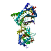 4nhkC 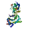 4nhmC 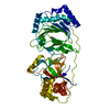 4nhxC 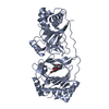 4nhyC 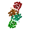 3kt7S  4nhn C: citing same article ( S: Starting model for refinement |
|---|---|
| Similar structure data |


-Links
 PDBj
PDBj

- Assembly
| Deposited unit | 
| ||||||||
|---|---|---|---|---|---|---|---|---|---|
| 1 |
| ||||||||
| Unit cell |
| ||||||||
| Components on special symmetry positions |
|
-Components
| #1: Protein | Mass: 74494.523 Da / Num. of mol.: 1 Source method: isolated from a genetically manipulated source Source: (gene. exp.) Strain: S288c / Gene: TPA1, YER049W / Plasmid: pNIC28 / Production host:  References: UniProt: P40032, Oxidoreductases; Acting on paired donors, with incorporation or reduction of molecular oxygen; With 2-oxoglutarate as one donor, and incorporation of one atom of oxygen into each donor |
|---|---|
| #2: Chemical | ChemComp-MN /   Mass: 54.938 Da / Num. of mol.: 1 / Source method: obtained synthetically / Formula: Mn Mass: 54.938 Da / Num. of mol.: 1 / Source method: obtained synthetically / Formula: Mn |
| #3: Chemical | ChemComp-OGA /   Mass: 147.086 Da / Num. of mol.: 1 / Source method: obtained synthetically / Formula: C4H5NO5 / Comment: inhibitor*YM Mass: 147.086 Da / Num. of mol.: 1 / Source method: obtained synthetically / Formula: C4H5NO5 / Comment: inhibitor*YM |
| #4: Water | ChemComp-HOH /  Mass: 18.015 Da / Num. of mol.: 74 / Source method: isolated from a natural source / Formula: H2O Mass: 18.015 Da / Num. of mol.: 74 / Source method: isolated from a natural source / Formula: H2O |
| Has protein modification | Y |
-Experimental details
-Experiment
| Experiment | Method: X-RAY DIFFRACTION / Number of used crystals: 1 |
|---|
- Sample preparation
| Crystal | Density Matthews: 2.6 Å3/Da / Density % sol: 52.72 % |
|---|---|
| Crystal grow | Temperature: 293 K / Method: vapor diffusion, sitting drop / pH: 7 Details: 0.8 mM MnCl2, 1.1 mM OGA, 0.1 M succinic acid, 12% PEG 3350, vapor diffusion sitting drop, temperature 293K, pH 7.0, VAPOR DIFFUSION, SITTING DROP |
-Data collection
| Diffraction | Mean temperature: 100 K | ||||||||||||||||||||||||||||||||||||||||||||||||||||||||||||||||||
|---|---|---|---|---|---|---|---|---|---|---|---|---|---|---|---|---|---|---|---|---|---|---|---|---|---|---|---|---|---|---|---|---|---|---|---|---|---|---|---|---|---|---|---|---|---|---|---|---|---|---|---|---|---|---|---|---|---|---|---|---|---|---|---|---|---|---|---|
| Diffraction source | Source: ROTATING ANODE / Type: RIGAKU FR-E+ SUPERBRIGHT / Wavelength: 1.5418 Å | ||||||||||||||||||||||||||||||||||||||||||||||||||||||||||||||||||
| Detector | Type: RIGAKU SATURN 944+ / Detector: CCD / Date: Jun 7, 2013 | ||||||||||||||||||||||||||||||||||||||||||||||||||||||||||||||||||
| Radiation | Protocol: SINGLE WAVELENGTH / Monochromatic (M) / Laue (L): M / Scattering type: x-ray | ||||||||||||||||||||||||||||||||||||||||||||||||||||||||||||||||||
| Radiation wavelength | Wavelength: 1.5418 Å / Relative weight: 1 | ||||||||||||||||||||||||||||||||||||||||||||||||||||||||||||||||||
| Reflection | Resolution: 2.84→30 Å / Num. all: 18339 / Num. obs: 18332 / % possible obs: 99.96 % / Redundancy: 3.7 % / Biso Wilson estimate: 44.543 Å2 / Rmerge(I) obs: 0.178 / Χ2: 1.141 / Net I/σ(I): 5.6 | ||||||||||||||||||||||||||||||||||||||||||||||||||||||||||||||||||
| Reflection shell | Diffraction-ID: 1 / Redundancy: 3.7 %
|
-Phasing
| Phasing | Method: molecular replacement |
|---|
- Processing
| Software |
| ||||||||||||||||||||||||||||
|---|---|---|---|---|---|---|---|---|---|---|---|---|---|---|---|---|---|---|---|---|---|---|---|---|---|---|---|---|---|
| Refinement | Method to determine structure: MOLECULAR REPLACEMENT Starting model: pdb entry 3KT7 Resolution: 2.84→29.7 Å / Occupancy max: 1 / Occupancy min: 0.5
| ||||||||||||||||||||||||||||
| Displacement parameters | Biso max: 84.96 Å2 / Biso mean: 42.6496 Å2 / Biso min: 26.35 Å2 | ||||||||||||||||||||||||||||
| Refinement step | Cycle: LAST / Resolution: 2.84→29.7 Å
| ||||||||||||||||||||||||||||
| Refine LS restraints |
| ||||||||||||||||||||||||||||
| LS refinement shell | Resolution: 2.83→2.9 Å
|
