 Movie
Movie Controller

[English] 日本語
 Yorodumi
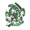
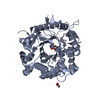
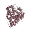
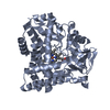
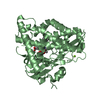
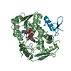
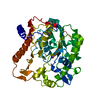
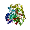
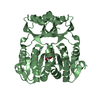
- PDB-4n48: Cap-specific mRNA (nucleoside-2'-O-)-methyltransferase 1 Protein ... -
+ Open data

- Basic information
| Entry | Database: PDB / ID: 4n48 | ||||||
|---|---|---|---|---|---|---|---|
| Title | Cap-specific mRNA (nucleoside-2'-O-)-methyltransferase 1 Protein in complex with capped RNA fragment | ||||||
 Components Components |
| ||||||
 Keywords Keywords | TRANSFERASE/RNA / methyltransferase / mRNA cap methylation / capped mRNA / TRANSFERASE-RNA complex | ||||||
| Function / homology |  Function and homology information Function and homology information7-methylguanosine mRNA capping / mRNA processing / methylation / methyltransferase cap1 / nucleic acid binding / methyltransferase cap1 activity / nucleoplasm / nucleus / cytoplasm Similarity search - Function | ||||||
| Biological species |  Homo sapiens (human) Homo sapiens (human) | ||||||
| Method |  X-RAY DIFFRACTION / SYNCHROTRON / MOLECULAR REPLACEMENT / Resolution: 2.704 Å X-RAY DIFFRACTION / SYNCHROTRON / MOLECULAR REPLACEMENT / Resolution: 2.704 Å | ||||||
 Authors Authors | Smietanski, M. / Werener, M. / Purta, E. / Kaminska, K.H. / Stepinski, J. / Darzynkiewicz, E. / Nowotny, M. / Bujnicki, J.M. | ||||||
 Citation Citation | Journal: Nat Commun / Year: 2014 Title: Structural analysis of human 2'-O-ribose methyltransferases involved in mRNA cap structure formation. Authors: Smietanski, M. / Werner, M. / Purta, E. / Kaminska, K.H. / Stepinski, J. / Darzynkiewicz, E. / Nowotny, M. / Bujnicki, J.M. | ||||||
| History |
|
- Structure visualization
| Structure viewer | Molecule: MolmilJmol/JSmol |
|---|
- Downloads & links
-Download
| PDBx/mmCIF format | 4n48.cif.gz | 181.4 KB | Display | PDBx/mmCIF format |
|---|---|---|---|---|
| PDB format | pdb4n48.ent.gz | 139.7 KB | Display | PDB format |
| PDBx/mmJSON format | 4n48.json.gz | Tree view | PDBx/mmJSON format | |
| Others |  Other downloads Other downloads |
-Validation report
| Arichive directory | https://data.pdbj.org/pub/pdb/validation_reports/n4/4n48ftp://data.pdbj.org/pub/pdb/validation_reports/n4/4n48 | HTTPS FTP |
|---|
-Related structure data
| Related structure data |  4n49SC  4n4aC S: Starting model for refinement C: citing same article ( |
|---|---|
| Similar structure data |

-Links
 PDBj
PDBj


- Assembly
| Deposited unit | 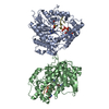
| ||||||||
|---|---|---|---|---|---|---|---|---|---|
| 1 | 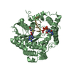
| ||||||||
| 2 | 
| ||||||||
| Unit cell |
|
-Components
| #1: Protein | Mass: 48935.754 Da / Num. of mol.: 2 Source method: isolated from a genetically manipulated source Source: (gene. exp.) Homo sapiens (human) / Gene: FTSJD2, KIAA0082, MTR1 / Production host:  #2: RNA chain | Mass: 1240.802 Da / Num. of mol.: 2 / Source method: obtained synthetically / Details: synthetic RNA #3: Chemical |   Mass: 398.437 Da / Num. of mol.: 2 / Source method: obtained synthetically / Formula: C15H22N6O5S Mass: 398.437 Da / Num. of mol.: 2 / Source method: obtained synthetically / Formula: C15H22N6O5S#4: Chemical | 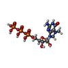  Mass: 539.223 Da / Num. of mol.: 2 / Source method: obtained synthetically / Formula: C11H20N5O14P3 Mass: 539.223 Da / Num. of mol.: 2 / Source method: obtained synthetically / Formula: C11H20N5O14P3#5: Water | ChemComp-HOH / |  Mass: 18.015 Da / Num. of mol.: 130 / Source method: isolated from a natural source / Formula: H2O Mass: 18.015 Da / Num. of mol.: 130 / Source method: isolated from a natural source / Formula: H2O |
|---|
-Experimental details
-Experiment
| Experiment | Method: X-RAY DIFFRACTION / Number of used crystals: 1 |
|---|
- Sample preparation
| Crystal | Density Matthews: 2.41 Å3/Da / Density % sol: 48.95 % |
|---|---|
| Crystal grow | Temperature: 291 K / Method: vapor diffusion / pH: 6.5 Details: 30% PEG 3350, 100 mM Bis-Tris [pH 6.5], and 100 mM NaBr, VAPOR DIFFUSION, temperature 291K |
-Data collection
| Diffraction | Mean temperature: 100 K |
|---|---|
| Diffraction source | Source: SYNCHROTRON / Site: BESSY  / Beamline: 14.1 / Wavelength: 0.91841 Å / Beamline: 14.1 / Wavelength: 0.91841 Å |
| Detector | Type: RAYONIX MX-225 / Detector: CCD / Date: Mar 13, 2012 |
| Radiation | Monochromator: Si-111 crystal / Protocol: SINGLE WAVELENGTH / Monochromatic (M) / Laue (L): M / Scattering type: x-ray |
| Radiation wavelength | Wavelength: 0.91841 Å / Relative weight: 1 |
| Reflection | Resolution: 2.7→30.334 Å / Num. all: 26308 / Num. obs: 24703 / % possible obs: 93.9 % / Observed criterion σ(F): 1.9 / Observed criterion σ(I): 1.9 |
| Reflection shell | Resolution: 2.7→2.86 Å / % possible all: 94.5 |
- Processing
| Software |
| ||||||||||||||||||||||||||||||||||||||||||||||||||||||||||||||||||||||
|---|---|---|---|---|---|---|---|---|---|---|---|---|---|---|---|---|---|---|---|---|---|---|---|---|---|---|---|---|---|---|---|---|---|---|---|---|---|---|---|---|---|---|---|---|---|---|---|---|---|---|---|---|---|---|---|---|---|---|---|---|---|---|---|---|---|---|---|---|---|---|---|
| Refinement | Method to determine structure: MOLECULAR REPLACEMENT Starting model: PDB ENTRY 4N49 Resolution: 2.704→30.334 Å / SU ML: 0.33 / σ(F): 2 / Phase error: 23.48 / Stereochemistry target values: ML
| ||||||||||||||||||||||||||||||||||||||||||||||||||||||||||||||||||||||
| Solvent computation | Shrinkage radii: 0.9 Å / VDW probe radii: 1.11 Å / Solvent model: FLAT BULK SOLVENT MODEL | ||||||||||||||||||||||||||||||||||||||||||||||||||||||||||||||||||||||
| Refinement step | Cycle: LAST / Resolution: 2.704→30.334 Å
| ||||||||||||||||||||||||||||||||||||||||||||||||||||||||||||||||||||||
| Refine LS restraints |
| ||||||||||||||||||||||||||||||||||||||||||||||||||||||||||||||||||||||
| LS refinement shell |
|
