 Movie
Movie Controller

[English] 日本語
 Yorodumi
- PDB-1y7p: 1.9 A Crystal Structure of a Protein of Unknown Function AF1403 f... -
+ Open data

- Basic information
| Entry | Database: PDB / ID: 1y7p | ||||||
|---|---|---|---|---|---|---|---|
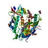
| Title | 1.9 A Crystal Structure of a Protein of Unknown Function AF1403 from Archaeoglobus fulgidus, Probable Metabolic Regulator | ||||||
 Components Components | Hypothetical protein AF1403 | ||||||
 Keywords Keywords | STRUCTURAL GENOMICS / UNKNOWN FUNCTION / Protein structure initiative / PSI / Archaeoglobus fulgidus / alpha-beta-alpha sandwich / Midwest Center for Structural Genomics / MCSG | ||||||
| Function / homology |  Function and homology information Function and homology informationHypothetical protein af1403; domain 2 / Uncharacterised conserved protein UCP006363, ACT-type / Domain of unknown function DUF5612 / Domain of unknown function (DUF5612) / : / AHAS small subunit-like ACT domain / ACT domain / ACT domain / ACT domain profile. / ACT domain ...Hypothetical protein af1403; domain 2 / Uncharacterised conserved protein UCP006363, ACT-type / Domain of unknown function DUF5612 / Domain of unknown function (DUF5612) / : / AHAS small subunit-like ACT domain / ACT domain / ACT domain / ACT domain profile. / ACT domain / ACT-like domain / CheY-like superfamily / Alpha-Beta Plaits / Rossmann fold / 2-Layer Sandwich / 3-Layer(aba) Sandwich / Alpha Beta Similarity search - Domain/homology | ||||||
| Biological species |   Archaeoglobus fulgidus (archaea) Archaeoglobus fulgidus (archaea) | ||||||
| Method |  X-RAY DIFFRACTION / SYNCHROTRON / SAD / Resolution: 1.9 Å X-RAY DIFFRACTION / SYNCHROTRON / SAD / Resolution: 1.9 Å | ||||||
 Authors Authors | Zhang, R. / Skarina, T. / Savchenko, A. / Edwards, A. / Joachimiak, A. / Midwest Center for Structural Genomics (MCSG) | ||||||
 Citation Citation | Journal: To be Published Title: 1.9A crystal structure of a hypothetical protein AF1403 from Archaeoglobus fulgidus Authors: Zhang, R. / Skarina, T. / Savchenko, A. / Edwards, A. / Joachimiak, A. / Midwest Center for Structural Genomics (MCSG) | ||||||
| History |
|
- Structure visualization
| Structure viewer | Molecule: MolmilJmol/JSmol |
|---|
- Downloads & links
-Download
| PDBx/mmCIF format | 1y7p.cif.gz | 136 KB | Display | PDBx/mmCIF format |
|---|---|---|---|---|
| PDB format | pdb1y7p.ent.gz | 108 KB | Display | PDB format |
| PDBx/mmJSON format | 1y7p.json.gz | Tree view | PDBx/mmJSON format | |
| Others |  Other downloads Other downloads |
-Validation report
| Arichive directory | https://data.pdbj.org/pub/pdb/validation_reports/y7/1y7pftp://data.pdbj.org/pub/pdb/validation_reports/y7/1y7p | HTTPS FTP |
|---|
-Related structure data
| Similar structure data | |
|---|---|
| Other databases |


-Links
 PDBj
PDBj


- Assembly
| Deposited unit | 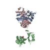
| ||||||||
|---|---|---|---|---|---|---|---|---|---|
| 1 | 
| ||||||||
| 2 | 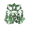
| ||||||||
| 3 | 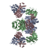
| ||||||||
| 4 | 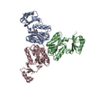
| ||||||||
| Unit cell |
| ||||||||
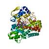
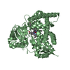
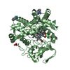
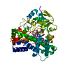
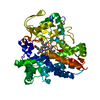
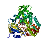
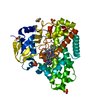
| Details | This protein existed as dimer. The deposited coords. of MolA and MolD form the biological dimer. |
-Components
| #1: Protein | Mass: 24454.412 Da / Num. of mol.: 3 Source method: isolated from a genetically manipulated source Source: (gene. exp.) Archaeoglobus fulgidus (archaea) / Strain: VC-16-DSM4304-ATCC49558 / Gene: AF1403 / Plasmid: pET15b / Species (production host): Escherichia coli / Production host:  #2: Sugar |   Type: D-saccharide, beta linking / Mass: 150.130 Da / Num. of mol.: 3 Type: D-saccharide, beta linking / Mass: 150.130 Da / Num. of mol.: 3Source method: isolated from a genetically manipulated source Formula: C5H10O5 #3: Chemical |   Mass: 65.409 Da / Num. of mol.: 3 / Source method: obtained synthetically / Formula: Zn Mass: 65.409 Da / Num. of mol.: 3 / Source method: obtained synthetically / Formula: Zn#4: Water | ChemComp-HOH / |  Mass: 18.015 Da / Num. of mol.: 234 / Source method: isolated from a natural source / Formula: H2O Mass: 18.015 Da / Num. of mol.: 234 / Source method: isolated from a natural source / Formula: H2O |
|---|
-Experimental details
-Experiment
| Experiment | Method: X-RAY DIFFRACTION / Number of used crystals: 1 |
|---|
- Sample preparation
| Crystal | Density Matthews: 1.958 Å3/Da / Density % sol: 35 % |
|---|---|
| Crystal grow | Temperature: 298 K / Method: vapor diffusion, hanging drop / pH: 8 Details: 0.2 M K/Na Tartr., 20% PEG3350, 0.05M Tris buffer, pH 8, VAPOR DIFFUSION, HANGING DROP, temperature 298K |
-Data collection
| Diffraction | Mean temperature: 100 K |
|---|---|
| Diffraction source | Source: SYNCHROTRON / Site: APS  / Beamline: 19-ID / Wavelength: 0.9798 Å / Beamline: 19-ID / Wavelength: 0.9798 Å |
| Detector | Type: SBC-2 / Detector: CCD / Date: Mar 7, 2004 / Details: mirrors |
| Radiation | Monochromator: Si 111 channel / Protocol: SINGLE WAVELENGTH / Monochromatic (M) / Laue (L): M / Scattering type: x-ray |
| Radiation wavelength | Wavelength: 0.9798 Å / Relative weight: 1 |
| Reflection | Resolution: 1.9→40 Å / Num. all: 117308 / Num. obs: 102527 / % possible obs: 87.4 % / Observed criterion σ(F): 2 / Observed criterion σ(I): 2 / Redundancy: 6.9 % / Biso Wilson estimate: 15.3 Å2 / Rmerge(I) obs: 0.09 / Net I/σ(I): 22.6 |
| Reflection shell | Resolution: 1.9→1.97 Å / Redundancy: 4.6 % / Rmerge(I) obs: 0.495 / Mean I/σ(I) obs: 1.534 / % possible all: 85.2 |
- Processing
| Software |
| ||||||||||||||||||||||||||||||||||||||||||||||||||||||||||||
|---|---|---|---|---|---|---|---|---|---|---|---|---|---|---|---|---|---|---|---|---|---|---|---|---|---|---|---|---|---|---|---|---|---|---|---|---|---|---|---|---|---|---|---|---|---|---|---|---|---|---|---|---|---|---|---|---|---|---|---|---|---|
| Refinement | Method to determine structure: SAD / Resolution: 1.9→36.34 Å / Rfactor Rfree error: 0.004 / Data cutoff high absF: 392525.79 / Data cutoff low absF: 0 / Isotropic thermal model: RESTRAINED / Cross valid method: THROUGHOUT / σ(F): 0 / Stereochemistry target values: Engh & Huber
| ||||||||||||||||||||||||||||||||||||||||||||||||||||||||||||
| Solvent computation | Solvent model: FLAT MODEL / Bsol: 65.6118 Å2 / ksol: 0.374876 e/Å3 | ||||||||||||||||||||||||||||||||||||||||||||||||||||||||||||
| Displacement parameters | Biso mean: 39 Å2
| ||||||||||||||||||||||||||||||||||||||||||||||||||||||||||||
| Refine analyze |
| ||||||||||||||||||||||||||||||||||||||||||||||||||||||||||||
| Refinement step | Cycle: LAST / Resolution: 1.9→36.34 Å
| ||||||||||||||||||||||||||||||||||||||||||||||||||||||||||||
| Refine LS restraints |
| ||||||||||||||||||||||||||||||||||||||||||||||||||||||||||||
| LS refinement shell | Resolution: 1.9→2.02 Å / Rfactor Rfree error: 0.012 / Total num. of bins used: 6
| ||||||||||||||||||||||||||||||||||||||||||||||||||||||||||||
| Xplor file |
|
