 Movie
Movie Controller

[English] 日本語
 Yorodumi
- PDB-4mos: Pyranose 2-oxidase H450G/V546C double mutant with 2-fluorinated g... -
+ Open data

- Basic information
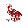
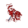
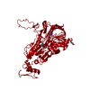
| Entry | Database: PDB / ID: 4mos | ||||||
|---|---|---|---|---|---|---|---|
| Title | Pyranose 2-oxidase H450G/V546C double mutant with 2-fluorinated galactose | ||||||
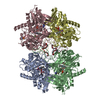
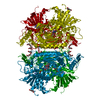
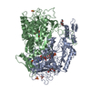
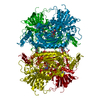
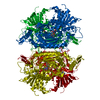
 Components Components | Pyranose 2-oxidase | ||||||
 Keywords Keywords | OXIDOREDUCTASE / GMC OXIDOREDUCTASE / PHBH FOLD / HOMOTETRAMER / FAD-BINDING / SUBSTRATE COMPLEX / FLAVINYLATION / INTRACELLULAR | ||||||
| Function / homology |  Function and homology information Function and homology informationpyranose oxidase / pyranose oxidase activity / flavin adenine dinucleotide binding / periplasmic space Similarity search - Function | ||||||
| Biological species |  Trametes ochracea (fungus) Trametes ochracea (fungus) | ||||||
| Method |  X-RAY DIFFRACTION / SYNCHROTRON / MOLECULAR REPLACEMENT / Resolution: 1.8 Å X-RAY DIFFRACTION / SYNCHROTRON / MOLECULAR REPLACEMENT / Resolution: 1.8 Å | ||||||
 Authors Authors | Tan, T.C. / Spadiut, O. / Gandini, R. / Haltrich, D. / Divne, C. | ||||||
 Citation Citation | Journal: Plos One / Year: 2014 Title: Structural Basis for Binding of Fluorinated Glucose and Galactose to Trametes multicolor Pyranose 2-Oxidase Variants with Improved Galactose Conversion. Authors: Tan, T.C. / Spadiut, O. / Gandini, R. / Haltrich, D. / Divne, C. | ||||||
| History |
|
- Structure visualization
| Structure viewer | Molecule: MolmilJmol/JSmol |
|---|
- Downloads & links
-Download
| PDBx/mmCIF format | 4mos.cif.gz | 245.6 KB | Display | PDBx/mmCIF format |
|---|---|---|---|---|
| PDB format | pdb4mos.ent.gz | 195.4 KB | Display | PDB format |
| PDBx/mmJSON format | 4mos.json.gz | Tree view | PDBx/mmJSON format | |
| Others |  Other downloads Other downloads |
-Validation report
| Arichive directory | https://data.pdbj.org/pub/pdb/validation_reports/mo/4mosftp://data.pdbj.org/pub/pdb/validation_reports/mo/4mos | HTTPS FTP |
|---|
-Related structure data
| Related structure data |  4moeC  4mofC  4mogC  4mohC  4moiC  4mojC  4mokC  4molC  4momC  4mooC  4mopC  4moqC  4morC C: citing same article ( |
|---|---|
| Similar structure data |


-Links
 PDBj
PDBj
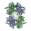
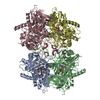
- Assembly
| Deposited unit | 
| ||||||||
|---|---|---|---|---|---|---|---|---|---|
| 1 | 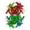
| ||||||||
| Unit cell |
|
-Components
-Protein , 1 types, 1 molecules A
| #1: Protein | Mass: 70476.969 Da / Num. of mol.: 1 / Mutation: H450G, V546C Source method: isolated from a genetically manipulated source Source: (gene. exp.) Trametes ochracea (fungus) / Gene: p2o / Plasmid: pET21(d+) / Production host:  |
|---|
-Sugars , 2 types, 2 molecules 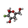
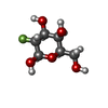
| #3: Sugar | ChemComp-GAF /  Type: D-saccharide, alpha linking / Mass: 182.147 Da / Num. of mol.: 1 / Source method: obtained synthetically / Formula: C6H11FO5 Type: D-saccharide, alpha linking / Mass: 182.147 Da / Num. of mol.: 1 / Source method: obtained synthetically / Formula: C6H11FO5 |
|---|---|
| #4: Sugar | ChemComp-2FG /  Type: D-saccharide, beta linking / Mass: 182.147 Da / Num. of mol.: 1 Type: D-saccharide, beta linking / Mass: 182.147 Da / Num. of mol.: 1Source method: isolated from a genetically manipulated source Formula: C6H11FO5 |
-Non-polymers , 3 types, 407 molecules 


| #2: Chemical | ChemComp-FDA /  Mass: 787.566 Da / Num. of mol.: 1 / Source method: obtained synthetically / Formula: C27H35N9O15P2 Mass: 787.566 Da / Num. of mol.: 1 / Source method: obtained synthetically / Formula: C27H35N9O15P2 |
|---|---|
| #5: Chemical | ChemComp-MES /  Mass: 195.237 Da / Num. of mol.: 1 / Source method: obtained synthetically / Formula: C6H13NO4S / Comment: pH buffer*YM Mass: 195.237 Da / Num. of mol.: 1 / Source method: obtained synthetically / Formula: C6H13NO4S / Comment: pH buffer*YM |
| #6: Water | ChemComp-HOH / Mass: 18.015 Da / Num. of mol.: 405 / Source method: isolated from a natural source / Formula: H2O |
-Details
| Has protein modification | Y |
|---|---|
| Sequence details | THIS IS A CLONING ARTIFACT |
-Experimental details
-Experiment
| Experiment | Method: X-RAY DIFFRACTION / Number of used crystals: 1 |
|---|
- Sample preparation
| Crystal | Density Matthews: 2.21 Å3/Da / Density % sol: 44.46 % |
|---|---|
| Crystal grow | Temperature: 298 K / Method: vapor diffusion, hanging drop / pH: 5.2 Details: 0.1 M MES, 50 mM MgCl2, 10% (w/v) monomethylether PEG 2000, pH 5.2, VAPOR DIFFUSION, HANGING DROP, temperature 298K |
-Data collection
| Diffraction | Mean temperature: 100 K |
|---|---|
| Diffraction source | Source: SYNCHROTRON / Site: Diamond  / Beamline: I24 / Wavelength: 0.9191 Å / Beamline: I24 / Wavelength: 0.9191 Å |
| Detector | Type: DECTRIS PILATUS 6M / Detector: PIXEL / Date: Feb 3, 2013 Details: Oxford Danfysik/SESO two stage demagnification using two K-B pairs of bimorph type mirrors |
| Radiation | Monochromator: ACCEL fixed-exit double-crystal Si(111) / Protocol: SINGLE WAVELENGTH / Monochromatic (M) / Laue (L): M / Scattering type: x-ray |
| Radiation wavelength | Wavelength: 0.9191 Å / Relative weight: 1 |
| Reflection | Resolution: 1.8→47.04 Å / Num. all: 59195 / Num. obs: 59195 / % possible obs: 100 % / Observed criterion σ(F): 0 / Observed criterion σ(I): 0 / Redundancy: 26.1 % / Net I/σ(I): 15.4 |
| Reflection shell | Resolution: 1.8→1.9 Å / Redundancy: 27.1 % / Mean I/σ(I) obs: 2.1 / % possible all: 100 |
- Processing
| Software |
| ||||||||||||||||||||||||||||||||||||||||||||||||||||||||||||||||||||||||||||||||||||||||||||||||||||||||||||||||||||||||||||||||||||||||||||||||||||||||||||||||||||||||||||||||||||||
|---|---|---|---|---|---|---|---|---|---|---|---|---|---|---|---|---|---|---|---|---|---|---|---|---|---|---|---|---|---|---|---|---|---|---|---|---|---|---|---|---|---|---|---|---|---|---|---|---|---|---|---|---|---|---|---|---|---|---|---|---|---|---|---|---|---|---|---|---|---|---|---|---|---|---|---|---|---|---|---|---|---|---|---|---|---|---|---|---|---|---|---|---|---|---|---|---|---|---|---|---|---|---|---|---|---|---|---|---|---|---|---|---|---|---|---|---|---|---|---|---|---|---|---|---|---|---|---|---|---|---|---|---|---|---|---|---|---|---|---|---|---|---|---|---|---|---|---|---|---|---|---|---|---|---|---|---|---|---|---|---|---|---|---|---|---|---|---|---|---|---|---|---|---|---|---|---|---|---|---|---|---|---|---|
| Refinement | Method to determine structure: MOLECULAR REPLACEMENT / Resolution: 1.8→47.04 Å / Cor.coef. Fo:Fc: 0.965 / Cor.coef. Fo:Fc free: 0.948 / SU B: 4.721 / SU ML: 0.076 / Cross valid method: THROUGHOUT / σ(F): 0 / ESU R: 0.106 / ESU R Free: 0.106 / Stereochemistry target values: MAXIMUM LIKELIHOOD / Details: HYDROGENS HAVE BEEN ADDED IN THE RIDING POSITIONS
| ||||||||||||||||||||||||||||||||||||||||||||||||||||||||||||||||||||||||||||||||||||||||||||||||||||||||||||||||||||||||||||||||||||||||||||||||||||||||||||||||||||||||||||||||||||||
| Solvent computation | Ion probe radii: 0.8 Å / Shrinkage radii: 0.8 Å / VDW probe radii: 1.2 Å / Solvent model: BABINET MODEL WITH MASK | ||||||||||||||||||||||||||||||||||||||||||||||||||||||||||||||||||||||||||||||||||||||||||||||||||||||||||||||||||||||||||||||||||||||||||||||||||||||||||||||||||||||||||||||||||||||
| Displacement parameters | Biso mean: 22.95 Å2
| ||||||||||||||||||||||||||||||||||||||||||||||||||||||||||||||||||||||||||||||||||||||||||||||||||||||||||||||||||||||||||||||||||||||||||||||||||||||||||||||||||||||||||||||||||||||
| Refinement step | Cycle: LAST / Resolution: 1.8→47.04 Å
| ||||||||||||||||||||||||||||||||||||||||||||||||||||||||||||||||||||||||||||||||||||||||||||||||||||||||||||||||||||||||||||||||||||||||||||||||||||||||||||||||||||||||||||||||||||||
| Refine LS restraints |
|