[English] 日本語
 Yorodumi
- PDB-4mm9: Crystal structure of LeuBAT (delta13 mutant) in complex with fluv... -
+ Open data
- Basic information
| Entry | Database: PDB / ID: 4mm9 | ||||||
|---|---|---|---|---|---|---|---|
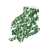
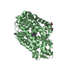
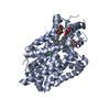
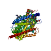
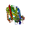
| Title | Crystal structure of LeuBAT (delta13 mutant) in complex with fluvoxamine | ||||||
 Components Components | Transporter | ||||||
 Keywords Keywords | TRANSPORT PROTEIN / transporter | ||||||
| Function / homology | Sodium:neurotransmitter symporter / Sodium:neurotransmitter symporter superfamily / Sodium:neurotransmitter symporter family / Sodium:neurotransmitter symporter family profile. / sodium ion transmembrane transport / plasma membrane / Fluvoxamine / Na(+):neurotransmitter symporter (Snf family) Function and homology information Function and homology information | ||||||
| Biological species |   Aquifex aeolicus (bacteria) Aquifex aeolicus (bacteria) | ||||||
| Method |  X-RAY DIFFRACTION / X-RAY DIFFRACTION /  SYNCHROTRON / SYNCHROTRON /  MOLECULAR REPLACEMENT / Resolution: 2.9 Å MOLECULAR REPLACEMENT / Resolution: 2.9 Å | ||||||
 Authors Authors | Wang, H. / Gouaux, E. | ||||||
 Citation Citation |  Journal: Nature / Year: 2013 Journal: Nature / Year: 2013Title: Structural basis for action by diverse antidepressants on biogenic amine transporters. Authors: Wang, H. / Goehring, A. / Wang, K.H. / Penmatsa, A. / Ressler, R. / Gouaux, E. | ||||||
| History |
|
- Structure visualization
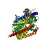
| Structure viewer | Molecule:  Molmil Molmil Jmol/JSmol Jmol/JSmol |
|---|
- Downloads & links
- Download
| PDBx/mmCIF format |  4mm9.cif.gz 4mm9.cif.gz | 112.3 KB | Display |  PDBx/mmCIF format PDBx/mmCIF format |
|---|---|---|---|---|
| PDB format |  pdb4mm9.ent.gz pdb4mm9.ent.gz | 86.5 KB | Display |  PDB format PDB format |
| PDBx/mmJSON format |  4mm9.json.gz 4mm9.json.gz | Tree view |  PDBx/mmJSON format PDBx/mmJSON format | |
| Others |  Other downloads Other downloads |
-Validation report
| Summary document |  4mm9_validation.pdf.gz 4mm9_validation.pdf.gz | 713.7 KB | Display |  wwPDB validaton report wwPDB validaton report |
|---|---|---|---|---|
| Full document |  4mm9_full_validation.pdf.gz 4mm9_full_validation.pdf.gz | 723.2 KB | Display | |
| Data in XML |  4mm9_validation.xml.gz 4mm9_validation.xml.gz | 19.1 KB | Display | |
| Data in CIF |  4mm9_validation.cif.gz 4mm9_validation.cif.gz | 25.3 KB | Display | |
| Arichive directory |  https://data.pdbj.org/pub/pdb/validation_reports/mm/4mm9 https://data.pdbj.org/pub/pdb/validation_reports/mm/4mm9 ftp://data.pdbj.org/pub/pdb/validation_reports/mm/4mm9 ftp://data.pdbj.org/pub/pdb/validation_reports/mm/4mm9 | HTTPS FTP |
-Related structure data
| Related structure data | 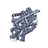 4mm4C 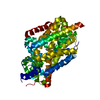 4mm5C 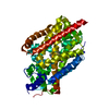 4mm6C 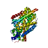 4mm7C  4mm8C  4mmaC  4mmbC  4mmcC 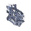 4mmdC 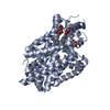 4mmeC 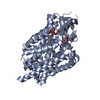 4mmfC C: citing same article ( |
|---|---|
| Similar structure data |
- Links
Links
- Assembly
| Deposited unit | 
| ||||||||
|---|---|---|---|---|---|---|---|---|---|
| 1 |
| ||||||||
| Unit cell |
|
- Components
| #1: Protein | Mass: 57988.266 Da / Num. of mol.: 1 Mutation: N21Y, G24D, I106S, T254S, S256G, A261V, I262L, Y265F, E290S, I359G, P362G, G408T, T409G Source method: isolated from a genetically manipulated source Source: (gene. exp.)   Aquifex aeolicus (bacteria) / Strain: VF5 / Gene: snf, aq_2077 / Production host: Aquifex aeolicus (bacteria) / Strain: VF5 / Gene: snf, aq_2077 / Production host:  | ||||
|---|---|---|---|---|---|
| #2: Chemical | | #3: Chemical | ChemComp-FVX / | #4: Water | ChemComp-HOH / | |
-Experimental details
-Experiment
| Experiment | Method:  X-RAY DIFFRACTION / Number of used crystals: 1 X-RAY DIFFRACTION / Number of used crystals: 1 |
|---|
- Sample preparation
| Crystal | Density Matthews: 2.8 Å3/Da / Density % sol: 56.08 % |
|---|---|
| Crystal grow | Temperature: 293 K / Method: vapor diffusion, hanging drop / pH: 7 Details: 100 mM NaPi, pH7.0, 100 mM NaCl, 32-34% PEG300, VAPOR DIFFUSION, HANGING DROP, temperature 293K |
-Data collection
| Diffraction | Mean temperature: 100 K |
|---|---|
| Diffraction source | Source:  SYNCHROTRON / Site: SYNCHROTRON / Site:  APS APS  / Beamline: 24-ID-E / Wavelength: 0.979 Å / Beamline: 24-ID-E / Wavelength: 0.979 Å |
| Detector | Type: ADSC QUANTUM 315 / Detector: CCD / Date: Feb 14, 2013 |
| Radiation | Monochromator: Cryogenically-cooled single crystal / Protocol: SINGLE WAVELENGTH / Monochromatic (M) / Laue (L): M / Scattering type: x-ray |
| Radiation wavelength | Wavelength: 0.979 Å / Relative weight: 1 |
| Reflection | Resolution: 2.9→51.3 Å / Num. all: 14312 / Num. obs: 14298 / % possible obs: 99.9 % / Observed criterion σ(I): -3 / Redundancy: 3.9 % / Rmerge(I) obs: 0.11 / Net I/σ(I): 5.2 |
| Reflection shell | Resolution: 2.9→3.08 Å / Redundancy: 4 % / Rmerge(I) obs: 0.55 / Mean I/σ(I) obs: 1.4 / % possible all: 100 |
- Processing
| Software |
| ||||||||||||||||||||||||||||||||||||||||||
|---|---|---|---|---|---|---|---|---|---|---|---|---|---|---|---|---|---|---|---|---|---|---|---|---|---|---|---|---|---|---|---|---|---|---|---|---|---|---|---|---|---|---|---|
| Refinement | Method to determine structure:  MOLECULAR REPLACEMENT / Resolution: 2.9→45.248 Å / SU ML: 0.43 / σ(F): 1.35 / Phase error: 36.12 / Stereochemistry target values: ML MOLECULAR REPLACEMENT / Resolution: 2.9→45.248 Å / SU ML: 0.43 / σ(F): 1.35 / Phase error: 36.12 / Stereochemistry target values: MLDetails: THE HIGH REAL SPACE R FOR THE LIGAND (FVX), IS DUE TO THE WEAK DENSITY OF TWO ALKYL CHAINS OF THE LIGAND (THE DENSITY FOR THE PHENYL GROUP IS VERY STRONG). THE AUTHORS MODELED THE LIGAND ...Details: THE HIGH REAL SPACE R FOR THE LIGAND (FVX), IS DUE TO THE WEAK DENSITY OF TWO ALKYL CHAINS OF THE LIGAND (THE DENSITY FOR THE PHENYL GROUP IS VERY STRONG). THE AUTHORS MODELED THE LIGAND POSITION BASED ON BOTH FO-FC AND 2FO-FC MAP. ALSO THE CURRENT POSITION AGREES WITH THE CHEMISTRY AND OTHER DRUGS POSITION THE AUTHORS DEPOSITED.
| ||||||||||||||||||||||||||||||||||||||||||
| Solvent computation | Shrinkage radii: 0.9 Å / VDW probe radii: 1.11 Å / Solvent model: FLAT BULK SOLVENT MODEL | ||||||||||||||||||||||||||||||||||||||||||
| Refinement step | Cycle: LAST / Resolution: 2.9→45.248 Å
| ||||||||||||||||||||||||||||||||||||||||||
| Refine LS restraints |
| ||||||||||||||||||||||||||||||||||||||||||
| LS refinement shell |
|
 Movie
Movie Controller



 PDBj
PDBj


